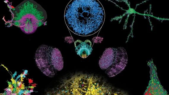
Novel microscopy method enables high-resolution study of neural brain circuits
A novel imaging technique combining two microscopy methods allowed researchers to visualize neural circuits across the brain at four-times that of typical resolutions, according to research published Jan. 17 in Science. The approach can be completed much faster than previously thought possible.
Combining expansion microscopy (ExM) with lattice light-sheet microscopy (LLSM), the team imaged entire fruit fly brains and the width of a mouse’s cortex in two to three days. Imaging the fly brain in color took 62.5 hours, compared to the years it would typically take using an electron microscope, according to the team.
“We’ve crossed a threshold in imaging performance,” corresponding author Eric Betzig, with Howard Hughes Medical Institute in Ashburn, Virginia, said in a news release. “That’s why we’re so excited. We’re not just scanning incrementally more brain tissue, we’re scanning entire brains.”
The microscopy technique required many processing steps. Two of the authors, Ruixuan Gao and Shoh Asano, both with the Massachusetts Institute of Technology’s MIT Media Lab in Cambridge, collected 50,000 cubes of data from the fruit fly brains. Then, members of their team analyzed the resulting 200 terabytes of data along with the 1,500 dendritic spines in mouse nerve cells, highlighting their neurons and counted all fly synapses. The results are clear images over large volumes in fractions of the time of normal microscopy methods, the team said in the release.
Their ultimate goal is to rapidly make maps of the entire nervous system, they added.
“The technology should enable statistically rich, large-scale studies of neural development, sexual dimorphism, degree of stereotypy, and structural correlations to behavior or neural activity, all with molecular contrast,” the authors concluded.

