Virtual reality proves a useful treatment tool for interventional radiologists
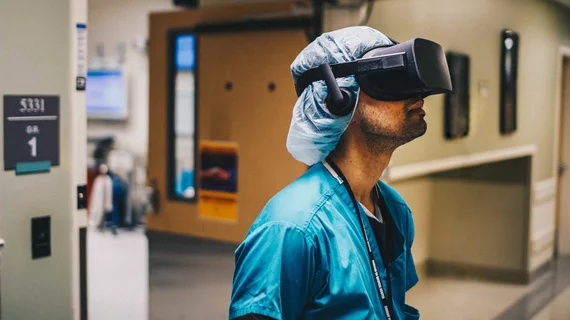
Research presented at this year’s Society of Interventional Radiology conference in Austin, Texas, suggests virtual reality (VR) could be useful to interventional radiologists, improving treatments using real-time 3D images from inside a patient’s vessels.
Wayne Monsky, MD, PhD, a professor of radiology at the University of Washington, and colleagues conducted a total of 18 simulated procedures for their study, which aimed to determine the feasibility of using a catheter to navigate a patient’s anatomy in real-time VR. The technique could be a novel way to improve radiologic treatments.
“Virtual reality will change how we look at a patient’s anatomy during an interventional radiology treatment,” Monsky said in a statement. “This technology will allow physicians to travel inside a patient’s body instead of relying solely on 2D, black-and-white images.”
Using a CT angiography scan, Monsky and his team developed a 3D-printed model and a holographic image of blood vessels in a patient’s abdomen and pelvis. The radiologists then guided catheters fitted with electromagnetic sensors through the 3D printed model while a tracking system projected the image from the catheter through a VR headset.
The team compared the time it took them to steer the catheter from the femoral artery to three different targeted vessels against the time it took to complete the same path using conventional fluoroscopic guidance. In all 18 procedures, the mean time it took to reach the targeted vessels was less when using VR over fluoroscopy—VR took 17.6 seconds to reach the first vessel compared to 70.3 seconds using fluoroscopy in the 3D model and 171.2 seconds in a real-life setting.
Monsky said the VR approach doesn’t only benefit radiologists themselves—who reported they felt more confident, precise and efficient when using VR—but extends that benefit to patients with poorer access to healthcare.
“Currently, the life-saving potential of interventional radiology is limited to hospitals and areas with the resources to invest in image-guided technology,” he said. “There are 3 billion people worldwide in rural areas who don’t have this access. This technology could allow for portability and accessibility so that these procedures are brought to rural areas using nothing more than a suitcase.”

