‘Very exciting development’: Hyperspectral imaging technique roots out hidden stomach cancers
A new imaging technique based on near-infrared technology can identify potentially cancerous tumors hidden deep under protective layers of muscle tissue.
Japanese researchers paired machine learning with their hyperspectral imaging technique to detect stomach tumors that are typically difficult to observe using standard methods, such as endoscopy. Using this new approach, they were 86% accurate at identifying gastrointestinal stromal tumors in a small group of tissue samples.
"This technique is a bit like x-rays; the idea is that you use electromagnetic radiation that can pass through the body to generate images of structures inside," Hiroshi Takemura, PhD, a biomedical researcher at Tokyo University of Science, said in a statement Tuesday. "The difference is that x-rays are at 0.01-10 nanometers, but near-infrared is at around 800-2500 nm. At that wavelength, near-infrared radiation makes tissues seem transparent in images. And these wavelengths are less harmful to the patient than even visible rays."
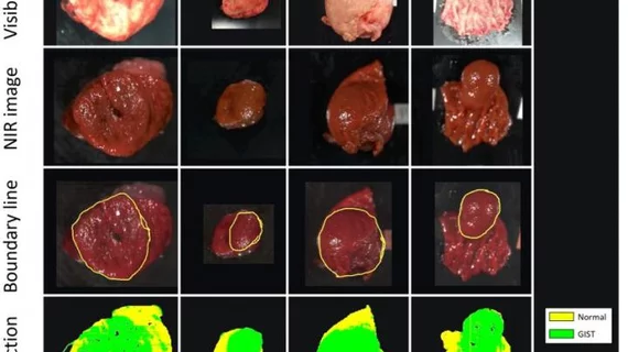
Takemura and his team performed near-infrared hyperspectral imaging on 12 surgically removed GIST tumors. A pathologist then analyzed the images to establish a border between healthy and tumor tissue. Those images were used to train the group’s machine learning algorithm.
The deep learning tool correctly color-coded tumor sections from non-tumor regions with 86% accuracy, even though 10 of the 12 abnormalities were completely or partly obscured by the innermost layer of stomach tissue.
"This is a very exciting development," Takemura explained. "Being able to accurately, quickly, and non-invasively diagnose different types of submucosal tumors without biopsies, a procedure that requires surgery, is much easier on both the patient and the physicians."
Going forward, the authors said they would like to improve their results using a larger training dataset and other tumor types. They are, however, currently working to perform NIR-HSI imaging on a patient instead of using surgically removed tissue.
Read the full study published in Scientific Reports here.

