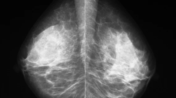
Machine learning model accurately predicts DCIS upstaging without invasive surgery
New machine learning models may be able to predict whether noninvasive cancers will eventually develop into more deadly, invasive cancers, according to new research published in Radiology.[1]
The study revealed that radiomic features in the mammograms of patients with ductal carcinoma in situ (DCIS) help predict upstaging, which would guide providers and patients in choosing the most effective treatment option.
DCIS is not life-threatening. However, patients with confirmed DCIS have an increased risk of eventually developing invasive ductal carcinoma. Currently, surgical excision is the standard of care for DCIS patients. Less-invasive alternatives are available, but they carry an increased risk of delayed detection of disease spread. Therefore, additional options are needed.
“Improving the presurgical diagnosis of occult invasive cancer in women with newly diagnosed DCIS can have important clinical implications and can assist providers and patients in choosing optimal management strategies,” corresponding author on the study, Eun-Sil Shelley Hwang, MD,, with the Department of Surgery at Duke University Medical Center, and co-authors explained.
For their research, experts used the mammograms of 700 women to create a machine learning model that could predict cancer upstaging. These scans were divided into a test set and a training set, and the team extracted 109 radiomic and four clinical features during the process.
Combining all clinical and radiomic features, the model used for the test set helped predict upstaging with an AUROC of 0.71. The upstaging rate was 16.3% (114 out of 700), while the model achieved 90% sensitivity and 92% negative predictive value.
The model that combined both radiomic and clinical features performed the best, the experts noted, but when judging the performance of one model alone, the radiomics model proved superior.
“We found that radiomic features derived from mammography can classify occult invasive disease in ductal carcinoma in situ, with performance superior to that of clinical criteria alone. This suggests potential to use imaging algorithms to improve patient care and assist in the selection of patients for clinical trials,” the authors concluded.
You can view the detailed research in Radiology.

