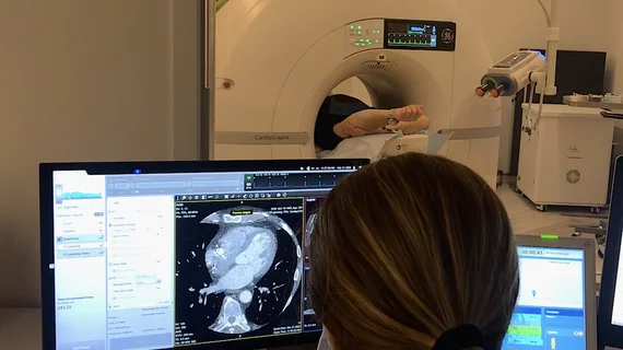
Fully automated CT body composition analysis predicts survival for CRC patients
A fully automated body composition analysis derived from CT imaging can be a valuable pretreatment tool for patients with colorectal cancer.
Previous research has suggested that manual or semi-automated body composition evaluations are beneficial for the risk stratification of patients with colorectal cancer. The authors of a new study published in the American Journal of Roentgenology recently found that a fully automated artificial intelligence-based analysis can also serve as a valuable predictive tool.
This came as the result of a retrospective analysis of 1,766 patients who had been diagnosed with colorectal cancer between January 2001 and September 2020. The researchers’ fully automated artificial intelligence-based algorithm was applied to portal venous-phase images from patients’ pretreatment scans in order to quantify the following: skeletal muscle attenuation at L3 (muscle attenuation), visceral adipose tissue (VAT) and subcutaneous adipose tissue (SAT) area at L3, and abdominal aorta Agatston score (aortic calcium). The findings were then compared to medical records to determine outcomes.
Using the fully automated body composition analysis, the experts were able to determine that patients with lower median muscle attenuation, SAT area and aortic calcium were less likely to survive. These risks were increased in patients in the lowest quartile for muscle attenuation and SAT area and in the highest quartile for aortic calcium. Conversely, they decreased for those in the highest quartile for VAT area and SAT area.
Clinical Impact
Corresponding author of the study Perry J. Pickhardt, MD, from the University of Wisconsin School of Medicine & Public Health, and colleagues maintained that their fully automated analysis could be a valuable prognostic tool used routinely in the future, stating that it “could improve initial risk stratification of patients with CRC.”
The study abstract can be viewed here.

