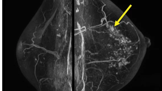
Surveillance breast MRI findings linked with future second cancer
Image features on breast MRIs of women with a history of breast cancer were recently linked with an increased risk of developing a secondary cancer in the future.
That’s according to new research focused specifically on associations between secondary breast cancers and background parenchymal enhancement—a known risk factor for breast cancer—on surveillance MRI exams. The study’s lead author Su Hyun Lee, MD, PhD, from the Department of Radiology at Seoul National University Hospital in Seoul, Korea, and colleagues detailed their findings in Radiology on Aug 30.
On contrast-enhanced breast MRI, background parenchymal enhancement (BPE) can vary in its appearance based on a number of factors, including prior radiation therapy treatments, hormonal status and breast density. While much is known about its appearance, the same cannot be said for its potential association with subsequent secondary cancers in breast cancer survivors.
To better understand this, Lee and colleagues conducted an analysis of 2,668 women who had undergone both breast MRI and surgery for primary breast cancer, none of whom had a prior diagnosis of secondary breast cancer. BPE was categorized as minimal, mild, moderate or marked and then compared to any tumor recurrences.
At a median follow-up of 5.8 years, 109 of the 2,668 women had developed a second breast cancer. The experts found that the women with secondary cancer displayed mild, moderate or marked BPE on imaging. Additionally, being younger in age (less than 45) at the time of the initial cancer diagnosis, being BRCA1/2 positive and having negative hormone receptor expression in the initial breast cancer were all independently associated with increased risks.
The authors suggested that their findings could impact the future of imaging surveillance for patients with a history of breast cancer, depending on whether additional risk factors are present.
“Our study results may help to stratify the risk of second breast cancer in women with a personal history of breast cancer and to establish personalized imaging surveillance strategies in terms of imaging modality and monitoring interval selection,” they explained.
In the future, the authors suggest that research should focus on monitoring changes in BPE on imaging completed before and after surgery and be compared to any subsequent cancers that may develop.
The study abstract can be viewed here.

