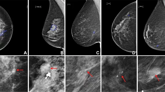
AI identifies breast lesion subtypes, could prevent unnecessary biopsies
Applying an artificial intelligence-based model to clinical mammograms could help identify not just breast cancer, but breast cancer subtypes as well.
Use of such a model was recently shown to have the potential to prevent up to 13% of unnecessary biopsies based on its performance in a cohort of nearly 3,000 women.
This and other findings were shared on November 1 in Radiology. Vesna Barros of IBM R&D Laboratories in Israel and colleagues explained how the use of artificial intelligence to identify lesion subtypes could benefit both clinics and patients.
“Although pathologic analysis remains the reference standard for breast lesion diagnosis, its invasive procedure can be a source of potential discomfort and anxiety,” the authors wrote. “Reducing the unnecessary biopsy rate can therefore considerably decrease health care costs and avoid unnecessary patient distress.”
For the study, algorithms were pretrained using imaging from more than 9,000 women who had at least one year of clinical and imaging history, in addition to biopsy-based histopathologic diagnoses. One model combined the use of convolutional neural networks with supervised learning algorithms and was trained on data from two sets of women—one set of 2,120 women in Israel and one set of 1,642 women in the United States.
The team assessed the models’ performances for differentiating between different lesion subtypes, including ductal carcinoma in situ, invasive carcinomas and benign lesions. In the Israeli group, the model achieved an average AUC of .88 and for the U.S. group, an AUC of .80 was observed.
A 99% sensitivity was recorded for the Israeli model. Experts noted that its use could have prevented 13% of unnecessary biopsies.
The authors suggested that the Israeli model might have performed better due to its larger test set. Additionally, they acknowledged the challenges associated with developing models that can identify lesion subtypes, as it requires large sets of data to reliably train them.
They noted that, in the future, the model could be enhanced by incorporating multi-modal imaging and genomic data, but that it could also have immediate benefits as-is for treatment planning purposes:
[O]ur model offers the potential to help radiologists recommend the appropriate follow-up approach on the basis of assessment of clinical, radiologic, and pathologic considerations.”
The study abstract can be viewed here.

