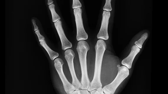
Has electronic cropping in digital radiography resulted in the death of collimation?
Collimating anatomy prior to image exposure is a known method of increasing image quality while also decreasing patients' radiation doses. However, in the era of digital radiography, it appears as though some techs are quite literally cutting corners when it comes to collimating.
How? By digitally cropping images at workstations after their exposures are made. While electronically cropping an image may seem like a harmless act in the grand scheme of things, the authors of a new paper published in the Journal of Medical Radiation Sciences explained that the habit is not without its consequences.
“On a digital image, lack of collimation has a detrimental effect on image quality, increasing the amount of scatter hitting the digital detector,” corresponding author Sally Ball of Princess Alexandra Hospital in Australia and colleagues wrote. “The increase in scatter can be a contributing factor to the histogram widening, resulting in a greyer image and a decreased spatial resolution, resulting in a lack of detail of the anatomy visualized.”
The experts sought to understand how common electronic cropping can be among radiographers, as well as what sort of impact, if any, a lack of collimation would have on final image quality. To do this, they compared measurements before and after electronic cropping was applied to the musculoskeletal radiographs of 359 patients to see if the exposed area was larger than a reference standard.
The experts focused on five projections—AP knee, AP shoulder, horizontal beam lateral hip, lateral cervical spine and lateral facial bones, resulting in a total of 1,071 measurements. They found that 61.2% of these measurements were above the agreed reference standard, meaning that the images were not collimated properly prior to image acquisition, but were instead digitally cropped afterwards.
Noting the increased workloads of radiographers, the experts suggested that digitally cropping images likely represents a time saver, adding that the digital age potentially “risks an environment in which patient throughput begins to take priority over image quality.”
However, the authors indicated that digital radiography also represents an opportunity for improvement via digital audits, which can be used to create targeted approaches to improving skills and image quality.
The detailed study can be viewed here.

