'Significant' brain abnormalities shown to persist 6 months after COVID recovery
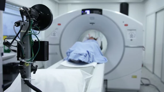
Using a specialized MRI technique, researchers recently uncovered ‘significant’ changes in the brains of COVID patients up to six months following their initial recovery.
The team utilized susceptibility-weighted imaging to assess how the virus impacted the brains of 46 COVID-recovered patients in comparison to 30 healthy controls with no history of ever contracting the respiratory infection. The regions that displayed the most significant differentiations in susceptibility values between the groups are known to be associated with neurological conditions such as fatigue, insomnia, anxiety, depression, headaches and other cognitive issues—all common complaints among many COVID long haulers.
Susceptibility-weighted imaging allows clinicians to understand how much certain materials, like blood, iron and calcium, etc., will become magnetized under a magnetic field. In turn, various neurologic conditions, such as stroke, microbleeds, malformations and other abnormalities are highlighted on imaging, explained co-author on the new study, Sapna Mishra, a PhD candidate at the Indian Institute of Technology in Delhi.
"Changes in susceptibility values of brain regions may be indicative of local compositional changes," Mishra said. "Susceptibilities may reflect the presence of abnormal quantities of paramagnetic compounds, whereas lower susceptibility could be caused by abnormalities like calcification or lack of paramagnetic molecules containing iron."
Experts found that the COVID group displayed significantly higher susceptibility values in comparison to the control group, with the findings most pronounced in the regions of the frontal lobe and brain stem.
The frontal lobe clusters consisted of abnormalities in the left and right orbital-inferior frontal gyrus and the adjacent white matter. Combined, these areas affect everything from language comprehension to attention, motor inhibition and imagery and social cognitive processes.
Additionally, the right ventral diencephalon region of the brain stem—an area that plays a significant role in regulating circadian rhythm, relaying sensory and motor skills and releasing hormones by way of the endocrine system—showed substantial differences in the COVID group of patients.
"This study points to serious long-term complications that may be caused by the coronavirus, even months after recovery from the infection," Mishra said. "The present findings are from the small temporal window. However, the longitudinal time points across a couple of years will elucidate if there exists any permanent change."
The research team is continuing their work with the same patient cohort to better understand the longitudinal nature of the changes observed in their study.
The details of this study will be presented at the 2022 Radiological Society of North America meeting being held in Chicago from November 27 to December 1.
To learn more, click here.

