New dual modality ultrasound technique offers greater 'accessibility, portability and cost effectiveness'
Researchers have developed a new dual modality imaging technique that offers super-resolution at a lower price point than standard imaging offerings.
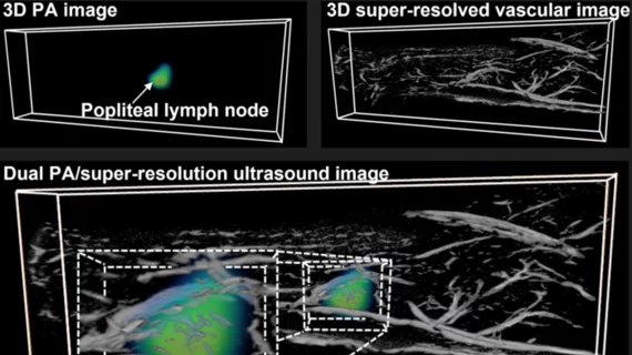
Developed by researchers at University of Illinois Urbana-Champaign, the technique integrates components of photoacoustic imaging and ultrasound localization microscopy with microbubbles. Researchers used a sparsity-constrained optimization method to combine the modalities and create the super-resolution imaging technique, which is capable of providing vascular and physiological imaging in less than 2 seconds per frame in vivo—a speed that reduces or eliminates the need for motion correction.
The hope is that the technique could lead to earlier detection of diseases by simultaneously identifying structural and functional abnormalities that standard ultrasound imaging methods alone cannot.
Experts involved in the new technique’s development detailed their work on April 18 in Nature Communications.
Corresponding author of the new paper Yun-Sheng Chen, with the Department of Electrical and Computer Engineering at the University of Illinois Urbana-Champaign, and colleagues suggested that not only does the super-resolution technique have the potential to facilitate earlier diagnosis, but it also offers “more accessibility, portability, and cost-effectiveness.”
“This technique is significantly cheaper than clinical medical imaging techniques, while providing similar functions,” the group noted.
The team demonstrated the technique’s effectiveness on mice for two challenging imaging scenarios for each respective modality alone: visualization of a dye-labeled lymph node showing nearby microvasculature, and kidney microangiography with tissue oxygenation. The technique allowed the team to visualize distinct vasculature while also giving them details on tissue functionality at the same time.
"The main benefit of this imaging is that it can provide multi-dimensional information,” the group explained. “We can add different layers of imaging. One layer can be structural, and another layer can be functional."
In the future, the team plans to explore the utility of incorporating molecular information into the technique.
The study abstract is available here.

