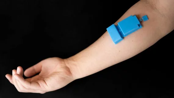
Wearable ultrasound device monitors muscle activity with great precision
A newly developed wearable ultrasound device could soon offer a more accurate alternative to electromyography (EMG) and pulmonary function exams.
Developed by researchers at the Aiiso Yufeng Li Family Department of Chemical and Nano Engineering at UC San Diego, the device can provide long-term, wireless monitoring of muscle activity and function. Experts are hopeful that it could have utility in multiple healthcare settings.
Details on its development and testing were shared Oct. 31 in Nature Electronics.
So far, the team has tested the device for two applications. First, it was applied to individuals’ rib cages to track the thickness and motion of their diaphragm. It was able to consistently and accurately record their breathing patterns. This is something that is often analyzed in patients on ventilators. What's more, in one small group, measures obtained through the device were used to accurately differentiate between individuals with and without chronic obstructive pulmonary disease.
Researchers are hopeful that it could be used in the future to both diagnose and monitor other respiratory conditions, co-author Joseph Wang, a distinguished professor in the Aiiso Yufeng Li Family Department of Chemical and Nano Engineering, said in a release.
“By tracking diaphragm activity, the technology could potentially support patients with respiratory conditions and those reliant on mechanical ventilation,” Wang suggested. “This technology could potentially be worn by individuals during their daily routines for continuous, long-term monitoring.”
The team has also used it to record muscle activity in extremities. When it is worn on the forearm, it can track activity in the wrist and the hand with great precision—perhaps even more accurately than an EMG could, the group suggested.
Armed with artificial intelligence, the device uses ultrasound signals to recognize hand and wrist motions and predict subsequent movements. The team recently used the device to control a robotic arm to pour water into beakers and play a video game. The group indicated that it could have great potential to improve prosthetics.
The device has three primary components: a single, highly sensitive ultrasound transducer; a custom wireless circuit that controls the transducer, records data and then wirelessly transmits it; and a lithium-polymer battery that lasts for a minimum of three hours.
Researchers are currently working on improving the device’s accuracy, portability and energy efficiency.

