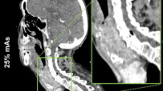
Deep learning denoising software can cut radiation exposure to 25% of conventional doses during neck CT
Deep learning denoising can reduce the amount of radiation patients are exposed to without sacrificing image quality of neck CT scans.
A new analysis in the European Journal of Radiology indicates that radiation dose levels at just 25% of conventional levels can be achieved with the help of deep learning denoising software. What’s more, the reconstruction technique did so while also improving subjective image quality, according to three radiologists tasked with rating the images.
The authors of the study suggested that the deep learning solution could help address multiple challenges related to the use of post-processing AI.
“The emergence of artificial intelligence-based post-processing denoising solutions presents a promising avenue for dose reduction in CT imaging,” corresponding author Andreas S. Brendlin, from the department of diagnostic and interventional radiology at Eberhard-Karls University in Germany, and co-authors wrote. “Integrating novel AI-based methods brings challenges, including potential losses in spatial information and blurring.”
For the study, researchers retrospectively examined neck CT scans completed between September and December 2023 on patients with suspected neck tumors. The team reconstructed the scans using Advanced Modeled Iterative Reconstruction (Original) at 100% and simulated 50% and 25% of conventional radiation doses, and post-processed using denoising software. Three blinded radiologists then rated each dataset based on image quality, diagnostic confidence, sharpness and contrast.
At each different dose level, significantly lower image noise and higher contrast-to-noise ratio (CNR) were observed for the denoised datasets in comparison to the original ones. The radiologists also rated image quality, diagnostic confidence, sharpness and contrast higher for denoising than for the original at 100 and 50 % of conventional doses.
“The subjective image quality decreased when the radiation dose was reduced in the original datasets, while the image quality remained high in the denoised datasets,” the authors explained. “Remarkably, no significant differences existed between the subjective image quality characteristics of the 25 % mAs denoised and 100% mAs original datasets.”
The authors suggested that similar algorithms could be utilized to minimize radiation exposure to all eligible patients, but especially in younger individuals who are at risk of radiation-induced malignancies after repeated scans.
The study abstract can be viewed here.

