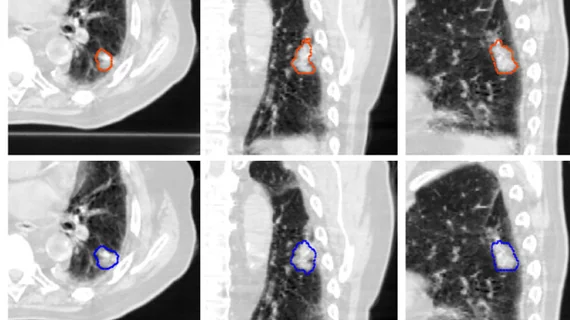
Deep learning model halves lung tumor segmentation times
Researchers have developed a near-expert-level deep learning model that accurately identifies and segments lung tumors on CT scans in significantly less time than radiologists.
While this is not the first model to take on lesion detection and segmentation successfully, this latest model has separated itself from others due to the especially large and diverse datasets on which it was tested and validated. Prior research has been hindered by small datasets and the need for manual inputs.
In a new study, the model was able to maintain its performance on scans completed using different types of CT scanners across multiple medical centers.
“To the best of our knowledge, our training dataset is the largest collection of CT scans and clinical tumor segmentations reported in the literature for constructing a lung tumor detection and segmentation model,” the study’s lead author, Mehr Kashyap, MD, resident physician in the Department of Medicine at Stanford University School of Medicine in California, said in a news release.
For the large-scale retrospective study, experts trained an ensemble 3D U-Net deep learning model on 1,504 CT scans with 1,828 segmented lung tumors. During testing, the model’s tumor volume predictions were compared to those of three radiologists based on measures of sensitivity, specificity, false positive rate and Dice similarity coefficient (DSC).
The model achieved 92% sensitivity and 82% specificity in detecting lung tumors, also yielding DSC results similar to the radiologists, at 0.77 compared to 0.80, respectively. Total segmentation time also was significantly shorter for the model in comparison to the human readers (mean 76.6 vs. 166.1–187.7 seconds)
The improvements could be owed to the use of a 3D U-Net architecture to develop the model, as if offers advantages over 2D architecture.
“By capturing rich interslice information, our 3D model is theoretically capable of identifying smaller lesions that 2D models may be unable to distinguish from structures such as blood vessels and airways,” Kashyap noted.
He added that the approach could have “wide-ranging implications” and positively assist providers in planning and managing lung cancer treatment.
Learn more about the findings here.

