New IR procedure could be a viable alternative to surgery for cubital tunnel syndrome
A new paper in the European Journal of Radiology details the potential of a newer minimally invasive ultrasound procedure as a viable alternative to surgery for cubital tunnel syndrome (CuTS).
Over time, constant compression of the ulnar nerve can cause chronic pain, numbness, weakness and even deformities in parts of the hand and wrist. Conservative treatments for the condition include physical therapy, over-the-counter anti-inflammatory medications and behavioral changes. However, if symptoms do not resolve and interfere with an individual’s day-to-day functioning, providers often turn to more invasive options, such as surgery or corticosteroid injections in the joint.
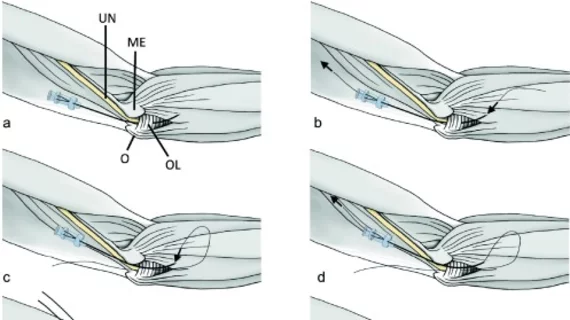
A newly developed ultrasound-guided thread technique (first described in Guo et al. and Kang et al.) could provide patients relief without the need for more invasive surgeries that are often accompanied by unpleasant side effects and prolonged downtime. The method involves guiding a thread around Osborne’s ligament under ultrasound guidance and then using small sawing motions to cut it. It takes around 20 minutes to perform.
“Due to the more invasive nature of surgical procedures, they are associated with greater intraoperative damage to surrounding tissue, extended rehabilitation periods, larger scars, and a heightened risk of recompression, in contrast to the minimally invasive ultrasound-guided thread release approach,” corresponding author Suren Jengojan, from the Department of Biomedical Imaging and Image-guided Therapy at the Medical University of Vienna, in Austria, and colleagues noted.
Researchers sought to delve deeper into the efficacy and safety of the procedure. To do this, they performed the ultrasound-guided release technique on 10 softly embalmed anatomic specimens. After the procedure, the team did a thorough examination of the anatomy surrounding Osborne’s ligament to check for any signs of damage it may have caused.
Encouragingly, there were no signs of damage in the area. The surrounding nerves, blood vessels, tendons and muscles all remained intact and unharmed, while the ligament was successfully cut in each cadaveric arm.
Although each procedure was successful, the group noted that the ultrasound quality varied, even with experienced radiologists completing the procedures.
“While proficient radiologists successfully executed the ligament transection without harm, it could prove more challenging for junior radiologists with limited ultrasound experience. Thus, a solid grasp of anatomical knowledge and adept handling of ultrasound, especially in an off-plane view, are essential prerequisites for the safe conduct of the procedure, with a requisite amount of practice,” the authors suggested.
Although all studies on the technique have been completed on cadavers thus far, the team maintained that it could still have a role as “an efficient and secure alternative to existing procedures.” They suggested that future studies assess the safety and efficacy in living patients and focus on any subsequent side effects that may emerge, including post-release instability, as those cannot be monitored in cadavers.
Learn more about the procedure here.

