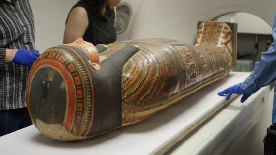
Mobile CT unit helps Field Museum experts gain new insight into ancient history
Researchers at the Field Museum of Chicago are using CT scans to unlock mysteries of the past lives of mummified people.
The team has been utilizing a mobile unit parked outside the museum to study the mummies in a more sensitive, person-centered approach that helps to preserve their wrappings. Over a four-day period during the summer, researchers scanned 26 mummified individuals from the museum. Through this, they have been able to answer questions related to the ages, lifestyles and even causes of death of the individuals scanned.
“From an archeological perspective, it is incredibly rare that you get to investigate or view history from the perspective of a single individual,” Stacy Drake, human remains collection manager at the Field Museum, said in a release on the project. “This is a really great way for us to look at who these people were—not just the stuff that they made and the stories that we have concocted about them, but the actual individuals that were living at this time.”
So far, the scans have helped researchers gain new insights into the lives of two of the museum’s most famous mummies— Lady Chenet-aa and Harwa.
For years, experts have been stumped by how exactly Lady Chenet-aa got into her cartonnage, as the funerary box did not appear to contain a point of entry. Thanks to the CT scans, researchers were able to identify a posterior seam down the backside of Chenet-aa's wrappings. This enabled the team to determine how the mummy was preserved and laid to rest.
The scans also provided new information on how old she was when she died, what sort of foods she ate and what she valued. For example, imaging showed that supplementary eyes had been placed in her eye sockets. JP Brown, senior conservator of anthropology at Field, explained that the decision to have the eyes placed was very intentional for Chenet-aa.
“The ancient Egyptian view of the afterlife is similar to our ideas about retirement savings. It's something you prepare for, put money aside for all the way through your life, and hope you've got enough at the end to really enjoy yourself,” Brown said. “The additions are very literal. If you want eyes, then there needs to be physical eyes, or at least some physical allusion to eyes.”
New information on one of the museum’s most popular mummies, Harwa, also was unearthed using CT. Harwa is famous for being the first mummified person to fly in an airplane in 1939; he also was the first of his kind to be lost in luggage when being transported from the New York World’s Fair.
The new scans of Harwa provided detailed information on the type of life he lived. Experts were able to determine that he lived into his 40s, which was no small feat 3,000 years ago. They also were able to ascertain that Harwa likely lived a fairly easy life, as his imaging showed no indications that his body had been subjected to hard physical labor. What’s more, his teeth were in excellent condition, suggesting that he had access to more high-quality foods.
Officials at Field have emphasized the importance of this new approach to studying mummified remains in a way that is dignified and respectful to the deceased subjects. The project is set to continue through 2025.
Learn more here.

