
Deep learning model enables routine radiographs to be used for osteoporosis screening
Deep learning techniques could open new doors for screening patients vulnerable to major fractures through routine X-ray imaging.
Currently, the gold standard for identifying low bone mineral density (BMD) is dual-energy X-ray absorptometry (DXA). However, many individuals with low BMD are unaware of their condition because they fail to get screened through DXA or are ineligible due to age, leaving many at-risk patients to fall through the cracks. These patients are at an increased risk of sustaining major fractures that could be debilitating and even deadly in some cases.
New research published in the European Journal of Radiology suggests that, with the help of a deep learning (DL) model, routine radiographs of the foot and ankle could be used to identify patients at risk of osteopenia or osteoporosis. Experts involved in the analysis believe their findings highlight a promising role for DL in preventing fractures prior to low BMD diagnosis.
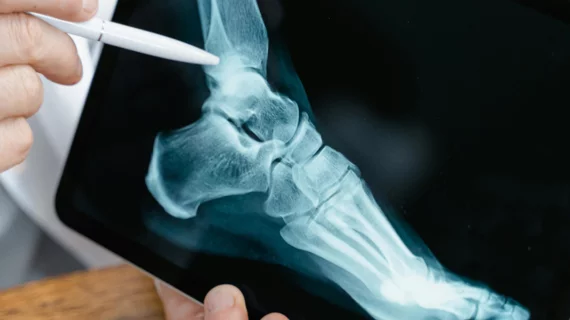
“The calcaneus is an important bone for estimating BMD. Radiographs of the foot and ankle may therefore be a reasonable choice to screen for low BMD,” explain co-authors Farid Gharehmohammadi and Ronnie Sebro, both with the Department of Radiology at MD Anderson Cancer Center in Houston. “These radiographs present a unique opportunity to identify individuals with low BMD (osteoporosis/osteopenia).”
Current radiology residency programs do not teach trainees how to assess BMD via radiographs. This, coupled with varying imaging techniques that create variability in image quality, could make it difficult to achieve consistent estimations of BMD across readers with different experience levels. This issue is what led the team to create their DL model.
The model was trained and validated on a set of more than 3,000 radiographs of patients who had undergone both routine X-rays of the foot/ankle and DXA scans within a 12-month period. Using a customized architecture, the model extracted several imaging features from the radiographs to predict whether patients would develop low BMD. The model’s predictions were then compared alongside patients’ DXA scans to measure performance.
In the test dataset, the model performed well, achieving an AUC of 0.87, sensitivity of 89.9%, specificity of 83.7%, positive predictive value of 90.8% and negative predictive value of 74.1%, respectively. It had an accuracy of 94.7% in the training/validation dataset and 89.9% in the test datasets, suggesting that it has the potential to accurately identify patients with osteopenia/osteoporosis using radiographs alone.
Its performance was consistent even when presented with different projections, medical conditions (i.e., arthritis) and imaging protocols. What’s more, its accuracy was maintained even when exams were completed using data from multiple different radiograph manufacturers and models.
“This discovery represents an advance that has the potential to increase orthopedists’, podiatrists’, rheumatologists’, and radiologists’ ability to identify patients who should go on to be screened for low BMD with DXA even before these patients would be eligible for BMD screening at the age recommended by United States Preventive Services Taskforce,” the group suggested. “This model will allow screening of younger individuals aged 50 and older without knowledge of their clinical risk profile for low BMD.”
The team is optimistic their model could prevent many individuals from developing osteoporosis and osteopenia by enabling providers to determine their risk earlier and initiate preventive treatments.
Learn more about the findings here.

In addition to her background in journalism, Hannah also has patient-facing experience in clinical settings, having spent more than 12 years working as a registered rad tech. She began covering the medical imaging industry for Innovate Healthcare in 2021.