Diamonds may be a sparkling method to reduce costs of medical imaging, drug studies
A new discovery involving diamonds may significantly cut costs related to medical imaging and drug-discovery devices, according to a team of researchers led by the U.S. Department of Energy and the University of California, Berkeley.
Researchers, led by Ashok Ajoy, PhD, a postdoctoral scholar at UC Berkeley, have discovered how to exploit defects in nanoscale and microscale diamonds to enhance MRI and nuclear magnetic resonance (NMR) system sensitivity. This development could, according to a May 18 press release, eliminate the need for costly, bulky magnets.
"Researchers will also seek to transfer this special tuning, known as spin polarization, to a harmless fluid such as water, and to inject the fluid into a patient for faster MRI scans," according to the press release. "Enhancing this spin polarization in the electrons of the diamonds’ atoms can be likened to aligning some compass needles pointing in many different directions to the same direction. These 'hyperpolarized' spins could provide a sharper contrast for imaging than conventional superconducting magnets."
The researchers used a green laser on microscale diamonds to subject them to a weak magnetic field. The team then used a microwave source on the diamonds to enhance the controllable spin polarization property, according to the researchers.
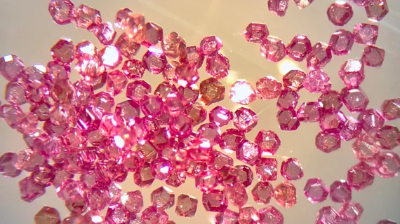
With a tool developed by Emanuel Druga, an electrician in the UC, Berkeley, the researchers confirmed and fine-tuned the spin polarization properties of the pinkish diamond samples that performed the best for testing, measuring one to five millionths of an inch across.
The diamonds, which can be made economically by converting graphite, may be able to reduce medical imaging and drug-discovery tools in the future, according to the release.
The study was published online May 18 in the journal Science Advances and funded by the National Science Foundation and the Research Corporation for Science Advancement.

