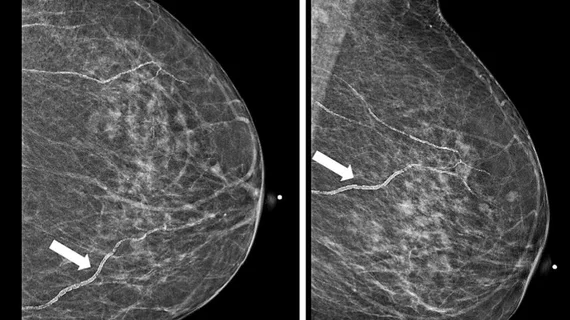
Arrows illustrate breast arterial calcifications for a 65-year-old woman on a screening mammography image (mediolateral oblique and craniocaudal views). Images and caption courtesy of Clinical Imaging.
There are no standards requiring radiologists to report on the presence of BACs, even though up to half of referring providers have indicated they would prefer to be made aware of the finding.
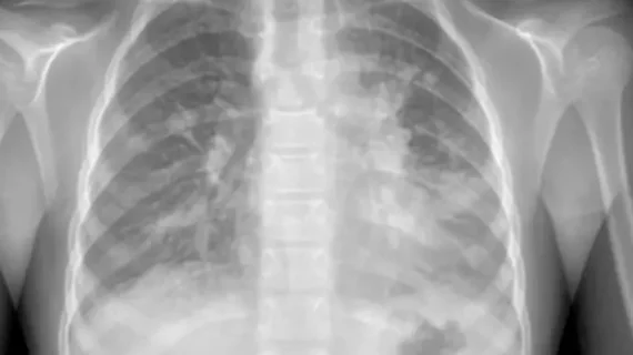
This chest X-ray image reveals areas of increased opacity in the lungs, indicative of an infection caused by Mycoplasma pneumoniae. Courtesy of Wikimedia Commons Image Source.
Following a recent surge of mycoplasma pneumoniae pneumonia cases, experts have issued new guidance to help providers quickly identify and treat the condition, with imaging playing a prominent role.
Despite the great progress that has been made toward the clinical implementation of AI, new data caution against trusting the technology as a single reader in certain settings.
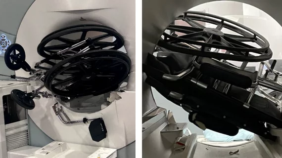
Isodose moderately hypofractionated radiotherapy allows patients to undergo just 4-5 weeks of treatment, compared to conventional therapy that can take around 8.
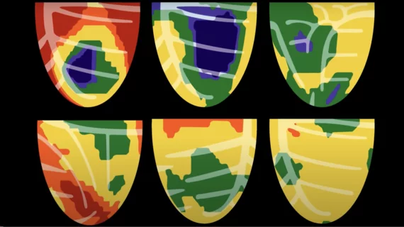
Clinicians have been using HeartSee to diagnose and treat coronary artery disease since the technology first debuted back in 2018. These latest updates, set to roll out to existing users, are designed to improve diagnostic performance and user access.