Trailblazers: iCAD’s groundbreaking tomosynthesis CAD solution uses deep learning to reduce reading times
The growing influence of artificial intelligence and deep learning in healthcare has led some writers to theorize that certain specialties, including radiology, would soon be “replaced” by machines. But Jeff Hoffmeister, MD, iCAD vice president and medical director, says such concerns couldn’t be further from the truth.
“When artificial intelligence first started to be used in radiology, I do think radiologists wondered if it was good enough and if it could end up completely replacing them,” Hoffmeister says. “But it’s been around long enough that I think they are starting to understand that it will not replace them. It may replace some of the more mundane, repetitive tasks radiologists do, but that empowers them to go out and focus on more complicated tasks a computer can’t do, and it should make their careers more interesting.”
iCAD’s PowerLook® Tomo Detection solution is a perfect example of how deep learning can improve patient care and empower radiologists. Digital breast tomosynthesis (DBT) continues to gain popularity among radiologists, yet scrolling through the hundreds and hundreds of images it produces can be tiresome and time-consuming. It typically takes a radiologist at least twice as long to read tomosynthesis compared to 2D mammography, which has created a certain level of apprehension to this superior technology.
“With tomosynthesis, radiologists can do a much better job at seeing cancers, but scrolling through all of those images just takes so much time that it’s hard to maintain concentration to say focused on the images,” Hoffmeister says. “It’s similar to a movie loop—as you’re scrolling through the images, you can focus on one area, but then you have to scroll back and forth a few times before you feel like you’ve adequately looked at the full set of images. That’s one of the biggest challenges with tomosynthesis, just being able to focus on multiple areas and the time it takes to do that.”
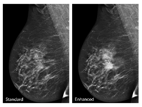
PowerLook® Tomo Detection, however, changes that dynamic. This new solution—which has received full FDA approval—is a powerful workflow tool that takes full advantage of the computer’s ability to “learn” the characteristics of cancer, scrolling through every image captured during tomosynthesis and extracting any suspicious lesions it finds. Those extractions are then blended into a synthetic 2D image, which Hoffmeister says is “much more natural for the radiologist to review.” The radiologist can then look at each suspicious area on the 2D image, and with one click of their mouse, they are taken to the actual slices for a more detailed examination.
“With PowerLook® Tomo Detection, readers get to the relevant areas much more quickly and then make the determination if what the computer found is something suspicious enough to warrant additional imaging evaluation or not,” Hoffmeister says. “We reduce the radiologist’s reading time with this technology without sacrificing their actual performance.”
In late 2015, iCAD conducted a clinical study to put the new solution’s performance to the test. Twenty radiologists read more than 200 cases with and without CAD, with a month of time in between to avoid memory bias. The results revealed that reading time had been reduced by an average of 29.2 percent, with no change in reader performance.
“With this study, we had two primary objectives: to show that radiologist performance is not inferior with CAD and to improve reading times,” Hoffmeister says. “We achieved both objectives. Sometimes, technology can be hard to demonstrate, but this is a powerful product that produces strong clinical data.”
PowerLook® Tomo Detection can provide radiology departments and private practices with more than just reduced reading times, allowing specialists to provide more care in any given day.
Hoffmeister also believes this solution can lead to increased overall receptiveness to digital breast tomosynthesis. While some specialists may be less than enthusiastic about current reimbursements for tomosynthesis, reducing reading times by nearly 30 percent allows that reimbursement to be much more aligned with the care being provided.
Looking ahead
Since it is the very nature of artificial intelligence to continuously learn new things and adapt to new data, the power and performance of PowerLook® Tomo Detection are sure to improve as time goes on.
“Higher-powered computers are becoming more available, and digital modalities are providing us with larger databases, so all of these things will come together to help deep learning do more and more for radiology in the next 5 to 10 years,” Hoffmeister says. “It’s a very exciting time to see how deep learning will impact these technologies and empower radiologists in the future.”
And if any radiologists are still concerned that this new technology will replace their job instead of helping them provide more high-quality care to more people, Hoffmeister advises they consider what happened when virtual colonoscopies were introduced in Wisconsin. By reducing some of the “unpleasantness” of traditional colonoscopies, these new procedures resulted in more patients being screened—which, of course, led to more work for the physicians treating those individuals.
“Computers and technologies can expand radiology,” Hoffmeister says. “I’m excited to see how that plays out.”

