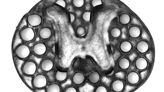
3D-printed implant created from MRIs may help treat spinal cord injuries
Using 3D printing, researchers from the University of California San Diego created spinal cord implants modeled from MRI scans that support nerve cell growth in spinal cord injuries and help restore lost physical mobility.
Detailed in a study published online Jan. 14 in Nature Medicine, the researchers found their four-centimeter-sized implants could support tissue regrowth, stem cell survival and the expansion of neural stem cell axons—long extensions on nerve cells that connect to other cells—from the scaffolding of the implants into the injured spinal cords of immobile rats.
“The new work puts us even closer to real thing because the 3D scaffolding recapitulates the slender, bundled arrays of axons in the spinal cord,” co-first author Kobi Koffler, PhD, assistant project scientist, said in a prepared statement. “It helps organize regenerating axons to replicate the anatomy of the pre-injured spinal cord.”
With their colleagues, co-senior author Shaochen Chen, PhD, a professor of nanoengineering at UC San Diego, and co-first author Wei Zhu, PhD, a nanoengineering postdoctoral fellow in Chen’s research group, used bioprinting technology to create the implants in less than 10 minutes.
The implants—each twice the width of human hair—aligned regenerating axons from one end of the spinal cord injury to the other while the scaffolding of the implant kept the axons in order and helped them grow in the right direction to complete the spinal cord connection in the rats.
“This [study] marks another key step toward conducting clinical trials to repair spinal cord injuries in people,” Koffler said. “The scaffolding provides a stable, physical structure that supports consistent engraftment and survival of neural stem cells. It seems to shield grafted stem cells from the often toxic, inflammatory environment of a spinal cord injury and helps guide axons through the lesion site completely.”
After a few months of grafting the implants into areas of severe spinal cord injury in rats, the new spinal cord tissue regrew completely across the injury and connected severed ends of the host spinal cord.
The researchers also noted that the circulatory systems of the treated rats had penetrated inside the implants to form functioning networks of blood vessels, which helped the neural stem cells survive. Ultimately, the rats regained motor function in their hind legs.

