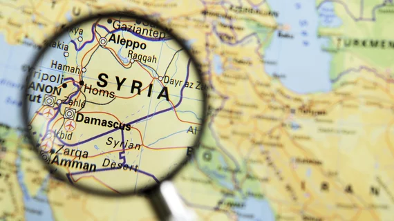
Social media promotes use of teleradiology by Syrian physicians
Social media has helped connect teleradiologists from around the world to the few physicians and imaging providers practicing in war-torn Syria, according to a study published May 31 in the Journal of the American College of Radiology.
Since the beginning of the Syrian Civil War in 2011, 485 medical facilities have been bombed and 841 medical personnel have been killed, wrote Abdulrahman Masrani, MD, from the Mallinckrodt Institute of Radiology at Washington University in St. Louis, and colleagues.
"The unique conflict ... has disrupted every aspect of the Syrians’ lives," the authors wrote. "It tremendously impacted the healthcare system with several reports of selective targeting of medical facilities and personnel."
Teleradiology interventions have been established to address a staggering number of casualties and traumatic injuries due to the conflict. Ghouta, a suburb located nine miles east of Damascus, has the highest death toll of medical personnel in Syria to date, wrote Masrani et al. The city's population dropped from 1.2 million to 400,000 people since a siege began 2013.
Some imaging equipment exists in Syrian field hospitals, including a few portable ultrasound machines, radiography machines, one functioning four-slice CT scanner and a dysfunctional CT scanner with confined field of view that is only used in select emergency cases, the authors wrote. However, they all lack MRI scanners and PACS, which has forced the only two radiologists to read directly from the scanner.
This shortage of on-site radiologists pushed the Syrian American Medical Society to establish the Teleradiology Relief Group (TRG) in February 2015, according to the authors. The TRG is comprised of four volunteer radiologists and two volunteer radiology residents. The group provides 24/7 coverage of imaging examinations through secret social media groups only accessible through mobile devices.
"Free and user-friendly social media platforms are feasible to utilize in providing humanitarian teleradiology services to areas under siege," the authors wrote. "Notwithstanding the limitations related to the siege, such projects are valuable in providing expert interpretation of CT, radiography and ultrasound examinations."
Using a satellite internet service, local doctors post to the Facebook group about their patients’ cases, medical images and lab results, if possible, the authors explained. The TRG teleradiology resident then comments on the post, providing an initial report and the attending radiologist approves it or not.
Since being established in 2015, the TRG has interpreted 497 radiological examinations, including 374 CTs, 199 plain films and four ultrasounds. Due to shortage of extremely experience contrast, 97 percent of CT scans were done without contrast.
"Even contrast-enhanced CT scans were of low diagnostic yield because of the poor-quality scanners, improper timing of intravenous administration, poor protocol and acquisition parameters, and lack of qualified technicians," according to the authors. "Repeating an examination was challenging because of the need for patient transport from the field hospital to the CT scanner facility."
Medical images are sent using DICOM format 57 percent of the time, and 43 percent of examinations were sent using JPEG format.
"Interpretation of JPEG images was difficult because of the inability to modify the window or level and the lack of proper sequence," the authors wrote. "In some instances, submitted images were taken by cell phone camera capturing poorly projected radiographic films or printed CT images on viewing boxes."
Nevertheless, preliminary reports are made within 24 hours after initially posting the exam in almost 90 percent of cases.

