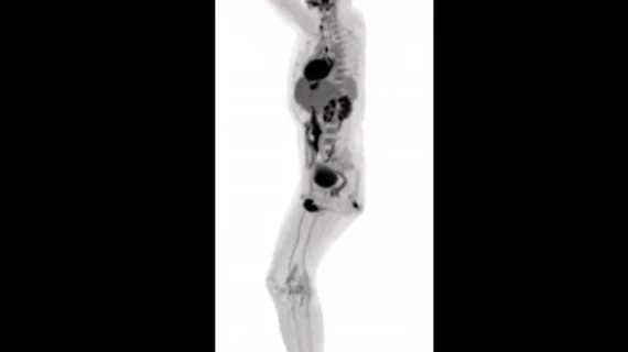
3D images from total-body scanner to be presented at RSNA 2018
Images from the world’s first whole-body MRI scanner are set to be presented at this year’s 2018 RSNA Annual Meeting in Chicago, according to a University of California, Davis statement.
The EXPLORER system, developed by Simon Cherry, PhD and Ramsey Badawi, PhD, both with UC Davis, is a combined PET and x-ray CT scanner that can image the entire body simultaneously. More efficient at capturing radiation, the scanner produces images in one second and can create videos tracking the movement of drugs through the body, according to a UC Davis press release.
"While I had imagined what the images would look like for years, nothing prepared me for the incredible detail we could see on that first scan," said Cherry, distinguished professor in the UC Davis Department of Biomedical Engineering, in the statement. "While there is still a lot of careful analysis to do, I think we already know that EXPLORER is delivering roughly what we had promised.”
Cherry believes the scanner could have great implications for clinical research and overall care due to its ability to produce higher-quality PET scans “than have ever been possible,” he said in the statement. EXPLORER requires 40-times less radiation than current methods, while producing whole-body scans in 20 to 30 seconds.
UC Davis is currently working with Shanghai-based United Imaging Healthcare (UIH)—which helped build the device—to install the system at the EXPLORER Imaging Center in Sacramento. The team hopes to begin using it for research in June 2019.

