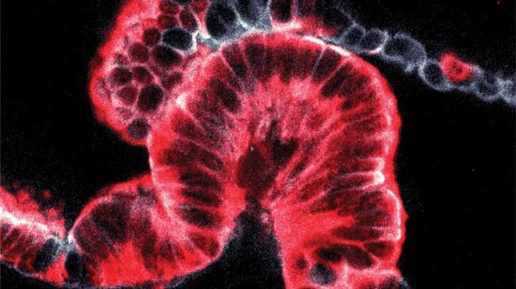
Novel 3D imaging method reveals origins of pancreatic cancer
A new three-dimensional (3D) imaging technique to analyze tissue samples allowed scientists to determine that pancreatic cancers can start and grow in two distinct ways, according to a Jan. 30 study published in Nature. The findings solve a question that’s plagued researchers for decades.
“To investigate the origins of pancreatic cancer, we spent six years developing a new method to analyze cancer biopsies in three dimensions,” said co-lead author, Hendrik Messal, PhD, from the Francis Crick Institute in London, in a news release. “This technique revealed that cancers develop in the duct walls and either grow inwards or outwards depending on the size of the duct. This explains the mysterious shape differences that we’ve been seeing in 2D slices for decades."
The pancreas relies on a system of ducts connected to other digestive organs, but the most common pancreatic cancers are found within the ducts, Messal et al. noted.
Using 3D imaging, the group found two types of cancer that originate in ductal cells: ‘endophytic’ tumors, which grow into the ducts, and ‘exophytic’ cancers, which grow outward. In an effort to determine why cancer cells grow a certain way the group created a simulation of the ducts using individual cell geometry to understand tissue shape, according to the authors.
They found the cancer grew outwards if the duct was less than approximately 20 micrometers—one-fifth of a millimeter. And after applying the 3D technique to other organs, Messal and colleagues found that cancers in the lung airways and ducts in the liver behave similarly to those in the pancreas.
“This technological breakthrough has the potential to unlock many unanswered questions of great importance in how we understand and treat pancreatic cancer,” said Andrew Biankin, Cancer Research UK’s pancreatic expert, in the release. “It’s crucial we better grasp how these cancers behave from the earliest stages, to help develop treatments for a disease where survival rates have remained stubbornly low.”

