
Advanced 3D imaging uncovers insights about prostate cancer aggressiveness
A new three-dimensional imaging technique can help physicians better understand the aggressiveness of prostate cancer and inform clinical decision making, according to a study shared in Cancer Research.
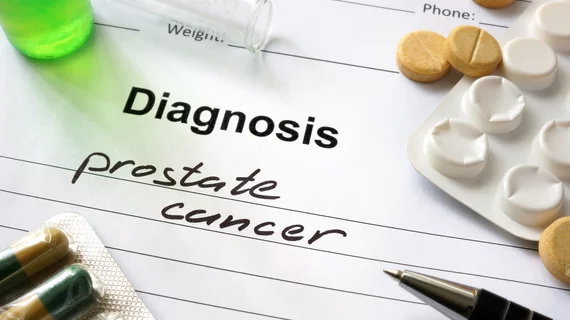
Prostate cancer is the most common form of the disease among men, but some cases are more time-sensitive than others. Analyzing abnormalities via traditional 2D methods, however, can make it difficult to diagnose such urgency.
University of Washington in Seattle scientists have created a “non-destructive” technique that images the entire 3D biopsy rather than just a slice. The enhanced features made it easier to spot cancers that were likely to recur within five years, according to the proof-of-principle results.
"This is exciting because it is the first of hopefully many clinical studies that will demonstrate the value of non-destructive 3D pathology for clinical decision making, such as determining which patients require aggressive treatments or which subsets of patients would respond best to certain drugs,” senior author Jonathan Liu, PhD, a professor of mechanical engineering and bioengineering at UW, explained Friday.
For their study, the team included 300 3D biopsies taken from 50 patients who underwent surgery more than a decade ago, affording them well-understood data on individual outcomes. Half of the biopsies included aggressive forms of cancer.
The 3D biopsies were stained to replicate methods used for 2D samples and imaged using the newly developed approach. Overall, the advanced imaging process uncovered additional details, including information regarding complex structures seen throughout cancerous tissue.
“This paper represents the first study in our lab to develop a novel computational pipeline to analyze our feature-rich datasets,” Liu explained. “As we continue to refine our imaging technologies and computational analysis methods, and as we perform larger clinical studies, we hope we can help transform the field of pathology to benefit many types of patients."
Read the full study in Cancer Research here.

Matt joined Chicago’s TriMed team in 2018 covering all areas of health imaging after two years reporting on the hospital field. He holds a bachelor’s in English from UIC, and enjoys a good cup of coffee and an interesting documentary.