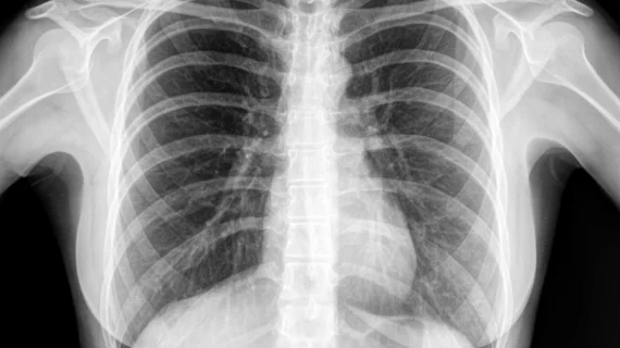
AI takes 10 seconds to diagnose pneumonia on chest x-rays
A new AI platform takes a mere 10 seconds to identify key findings on a patient’s chest x-ray, compared to the 20 minutes typically required.
The system—CheXpert—was developed using 188,000 chest images by researchers at Stanford University and fine-tuned by a team at Intermountain Medical Center to identify suspected pneumonia. If implemented in an emergency department, physicians may be able to treat patients sooner—vital to those suffering from pneumonia.
"CheXpert is going to be faster and as accurate as radiologists viewing the studies,” Nathan C. Dean, MD, principal investigator of the study, and section chief of pulmonary and critical care medicine at Intermountain Medical Center, said in a statement. “It's an exciting new way of thinking about diagnosing and treating patients to provide the very best care possible.”
Chest x-rays are commonly performed in patient with suspected pneumonia, according to the researchers. In an emergency department, it can take a while to get that image read which can result in delays in the start of an antibiotic treatment cycle for sick patients.
As part of their study, Intermountain researchers customized CheXpert on 6,973 images from its emergency department. They then had radiologists categorize chest x-rays from 461 patients as either “likely,” “likely-uncertain,” “unlikely-uncertain” or “unlikely” for pneumonia. The readers also identified scans thought to show pneumonia in many parts of the lungs and whether those patients had parapneumonic effusion.
Overall, CheXpert beat out radiologists at identifying key pneumonia findings. Additionally, in more than 50% of cases, the radiologists categorized patients differently. The AI model’s performance was “comparable” to these experts.
“In this initial study, we've demonstrated the algorithm's potential by validating it on patients in the emergency departments at Intermountain Healthcare,” Jeremy Irvin, PhD student at Stanford and research team member, said in the statement. “Our hope is that the algorithm can improve the quality of pneumonia care at Intermountain, from improving diagnostic accuracy to reducing time to diagnosis."
The study findings were presented Sept. 30 at the European Respiratory Society’s International Congress in Madrid.

