
AI algorithm reduces metal artifacts on CT imaging of metallic implants
Deep learning algorithms offer solutions to the issue of metal artifacts on CT scans of individuals with medical implants, new research suggests.
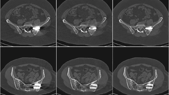
In comparison to uncorrected images and orthopedic metal artifact reduction (O-MAR)-corrected CT images, a deep learning-based metal artifact reduction technique (dl-MAR) offers superior image quality, according to newly published research in the European Journal of Radiology.
While the concept of utilizing deep learning to reduce artifacts on imaging is not a new one, prior studies of its potential were limited in their comparisons, authors of the new paper explained.
“Deep learning models trained on CT images with simulated metal artifacts have been shown to successfully reduce metal artifacts in images,” corresponding author Mark Selles, with the Department of Radiology and Nuclear Medicine at Amsterdam University in the Netherlands, and co-authors noted. “However, concerns about the generalizability of deep learning models stem from the simulation process of metal artifacts where metal implants are not always placed in anatomically correct positions, not all relevant physical effects are included, and only simulation of 2D CT slices is feasible.”
The team addressed this shortcoming by including comparisons between uncorrected and O-MAR-corrected images to DL-MAR-corrected images during both pre- and post-op exams of patients undergoing sacroiliac joint fusion. Six different regions of interest (ROIs) were placed on the metal implant and surrounding areas.
“A solid evaluation requires paired CT-images with and without metal, which only can be obtained by scanning a phantom or patients multiple times,” the group explained.
Both O-MAR and DL-MAR techniques resulted in improved image quality, but the deep learning algorithm yielded more significant artifact reduction in contralateral bone, the gluteus medius, contralateral gluteus medius, iliacus and contralateral iliacus.
This is the first study to use paired, real-world clinical CT images to evaluate DL-based artifact reduction techniques. The positive findings indicate real potential for such a technique’s use in the future, the team suggested, later describing the work as “a next step towards clinical application of deep learning-based metal artifact reduction techniques.
The study abstract is available here.

In addition to her background in journalism, Hannah also has patient-facing experience in clinical settings, having spent more than 12 years working as a registered rad tech. She began covering the medical imaging industry for Innovate Healthcare in 2021.