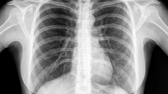
AI-assisted pneumothorax detection via chest radiographs reduces invasive procedures for patients
Identifying a pneumothorax on chest radiographs after patients undergo lung biopsy can be tricky, but new research published Tuesday suggests that a computer-aided detection (CAD) system could be key.
The most common complication of percutaneous transthoracic needle biopsy is pneumothorax, occurring after 16%-38% of procedures. Though most pneumothorax resolve with conservative treatment, some can be life-threatening and require more invasive interventions to prevent respiratory failure.
Chest radiographs are the standard for detecting pneumothorax, however, increased radiologist workloads can interfere with timely and precise detection.
“In clinical practice, prompt and accurate interpretation of radiographs is not always possible, resulting in a delayed diagnosis of pneumothorax,” corresponding author Chang Min Park, from the Department of Radiology at Seoul National University Hospital, South Korea, and co-authors explained.
Retrospective studies have shown deep learning algorithms accurately identify pneumothorax via chest X-rays. But like many studies pertaining to artificial intelligence applications, real-world accuracy has not yet been validated. This is what prompted the experts at Seoul National University Hospital to investigate the utility of CAD systems in clinical practice.
In 2020, the researchers implemented a CAD system for pneumothorax detection on chest radiographs at a single institution. They then compared the results of the CAD-assisted interpretations (completed February to November 2020) to previous reads performed before the system was applied (January 2018 to 2020).
Their results revealed that CAD-applied interpretations were superior in sensitivity, negative predictive values and accuracy (85.4%, 96.8%, 96.8%, respectively) compared to the non-CAD group (67.1%, 91.3%, 92.3%).
Encouragingly, the increased detection also resulted in fewer patients with pneumothorax requiring drainage catheter insertions. The experts impled that this could be due to the CAD’s ability to earlier detect pneumothorax. This would prompt providers to quickly initiate conservative treatments before the pneumothorax could progress to the point of becoming symptomatic, which would require more invasive treatments.
“We believe that the CAD system can help improve the safety of patients receiving lung biopsy and, furthermore, may be used to promptly detect and timely manage pneumothorax of any cause,” the experts suggested.
You can view the detailed study in Radiology.

