How AI-generated 'fake' X-rays can further medical research
The use of synthetic X-rays could help medical researchers to cut through the regulatory red tape that so often accompanies data sharing, especially within the realm of AI research, as it requires a substantial amount of data for training, testing and validation purposes.
But how reliable can synthetic radiologic images be in research settings? Are they as good as the real thing?
According to new work by researchers at the University of Jyväskylä's AI Hub Central Finland project, it is possible for synthetic medical images to be realistic enough to make specialized medical experts do a double take—a revelation that experts involved in the study suggested could eventually expedite the process of AI research.
“The use of synthetic data is not subject to the same data protection regulations as real data. Using synthetic data can facilitate collaboration between, for example, research groups, companies and educational institutions,” Sami Äyrämö, Head of Digital Health Intelligence Laboratory at the University of Jyväskylä, said in a news release.
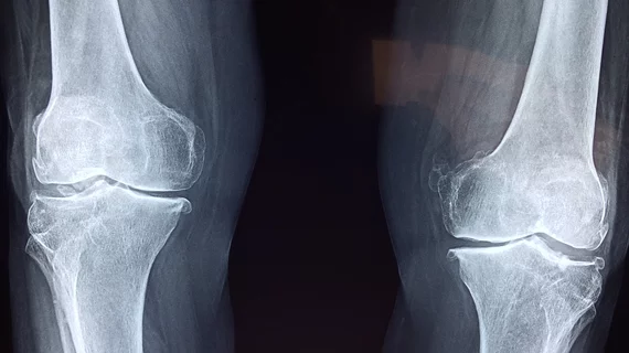
For the project, the team developed an artificial neural network to create synthetic knee X-ray images realistic enough that they could replace or complement real radiographic knee images for the sake of diagnosing and classifying osteoarthritis (OA).
Medical experts (15) were then asked to assess a group of images that included both real and synthetic knee X-rays in two phases—first to simply classify OA severity without being aware of the synthetic images, and then again to distinguish which images were real and which were fake.
The researchers found that, on average, the medical experts could not differentiate between the two sets of images.
These findings prompted experts involved in the study to suggest that synthetic data could help to address the issue of insufficient amounts of medical data required to conduct research, especially in situations when real patient data is limited.
“In addition, the neural network is capable of modifying synthetic X-ray images to expert specifications,” explained Fabi Prezja, the Doctoral Researcher responsible for developing the artificial neural network. “This capability is very powerful and allows potential future use for medical educational applications, and stress testing for other AI systems.”
To learn more, or to see some of the synthetic images, click here.

