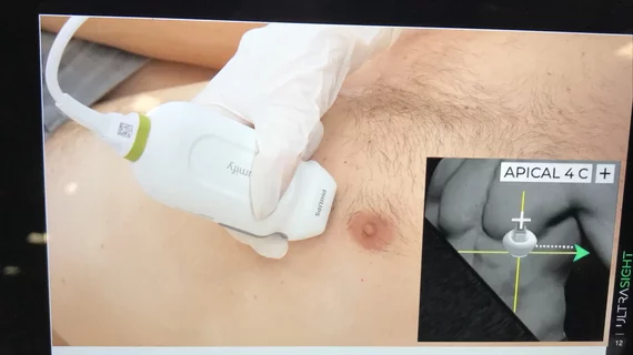
AI helps novice medical assistants perform high-quality echocardiograms
Novice medical assistants with no sonography experience were able to obtain diagnostic quality cardiac images with the help of AI, announced digital health company UltraSight at the Annual Conference of the Israel Heart Society in Tel Aviv.
In a blind pilot study, three medical assistants had eight hours of training and then conducted 2D transthoracic echocardiography (TTE) scans for 61 cardiac patients, guided in real time by AI software. Each patient was also scanned by a trained sonographer.
Three U.S. board-certified cardiologists then judged the resulting images to determine if they were of sufficient diagnostic quality to permit the assessment of left ventricular size and function, right ventricular size, and presence of significant pericardial effusion. All 61 scans from the novice medical assistants were judged to be diagnostic quality.
“These anatomical assessments are essential to determining heart pathologies,” said Roberto M. Lang, MD, director of the Noninvasive Cardiac Imaging Laboratory at the University of Chicago, in a statement about the study’s results. “This is crucial in critical care settings and emergency departments where decisions about cardiac treatment and care need to be made very quickly before conditions deteriorate. The fact that medical assistants were able to take such precise images and achieve near-perfect results with a handheld device is outstanding.”
The possibility for any medical professional to successfully capture diagnostic quality images can be helpful to speed up patient care in time-sensitive situations, especially in facilities where staffing is limited.
“Cardiac ultrasound is a powerful diagnostic tool enabling timely evaluation of a patient in the emergency department,” said UltraSight CEO Davidi Vortman. “However, its application is often limited by the availability of experienced users. This study is a significant step forward in making cardiac ultrasound more widely accessible to patients anywhere.”
Related Artificial Intelligence content:
AI-based mammo screening protocol reduces radiologist workload by 62%
Explainable AI model accurately auto-labels chest X-rays from open access datasets
AI model could open doors for greater access to obstetric ultrasound
AI cuts average CCTA reading time by 73%, helping radiologists detect coronary artery disease
