AI-generated synthetic brain MRI provides diverse, reliable training data
Researchers have created a deep-learning model that can generate synthetic brain MRIs to help train neural networks, according to research published in ArXiv. The technique may provide reliable, shareable training data.
“Data diversity is critical to success when training deep learning models,” wrote lead author Hoo-Chang Shin, with Nvidia Corporation, and colleagues. “Medical imaging data sets are often imbalanced as pathologic findings are generally rare, which introduces significant challenges when training deep learning models. We propose a method to generate synthetic abnormal MRI images with brain tumors by training a generative adversarial network (GAN).”
Scientists from Nvidia teamed up with Mayo Clinic in Rochester, Minnesota, and the MGH and BWH Center for Clinical Data Science in Boston for the project.
They trained the GAN using more than 3,400 MRIs contained in the Alzheimer’s Disease Neuroimaging Initiative data set, along with an additional 200 4D brain MRIs with tumors from the Multimodal Brain Tumor Image Segmentation Benchmark data set.
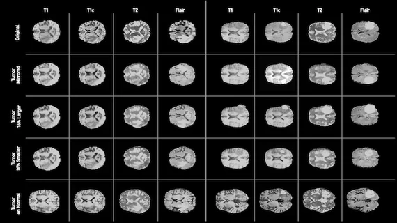
The GAN-generated images successfully produced synthetic abnormal brain tumor MRIs using image-to-image translation. Shin and colleagues found that by altering the GAN’s input label map, a more diverse set of data could be achieved.
“This offers an automatable, low-cost source of diverse data that can be used to supplement the training set,” the authors wrote. “For example, we can alter a tumor’s size, change its location, or place a tumor in an otherwise healthy brain, to systematically have the image and the corresponding annotation.”
Shin and colleagues noted the synthetic nature of the images means there are no patient data or privacy issues—allowing medical institutions to easily share the data they’ve generated.

