
AI tool predicts metabolic disease using 3D body scans
Combined with artificial intelligence software, 3D body scans provide valuable insight into whether a person is at risk of developing metabolic disease, a new study from researchers at Mayo Clinic indicates.
Metabolic syndrome is based on whether someone has three out of five of the following conditions: abdominal obesity, hypertension, high triglycerides, low HDL cholesterol and high fasting blood sugar. Individuals who have some form of metabolic disease are at heightened risk of heart attack, stroke, diabetes, liver disease and more.
These conditions develop slowly over time. Current body mass index (BMI) metrics and body composition scales are outdated, and do not consider the full picture of patients’ overall health. There need to be methods that take multiple personalized factors into account to more accurately predict who is at risk of serious metabolic decline, experts are finding.
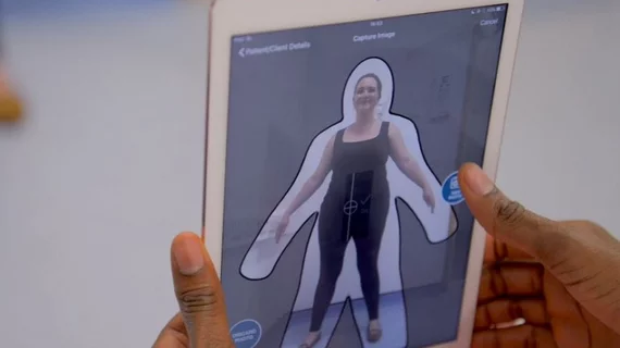
That’s where Mayo’s latest research comes into play. Scientists from the organization worked to develop an algorithm that incorporates information obtained during multi sensor white light 3D body volume scans (3D-BV) that collect over 600,000 linear data points. Information obtained from the scan is combined with patient demographics, blood tests and standard body measurements, in addition to front and side view photos to determine whether someone has or is at risk of developing metabolic issues.
"There is a need for a reliable, repeatable measure of metabolic syndrome risk and severity," Betsy Medina Inojosa, MD, a research fellow at Mayo Clinic and first author of the study, said in a release. "Body mass index measurements and bioimpedance scales that measure body fat and muscle are inaccurate for many people, and other types of scans are not widely available.”
The tool was developed and validated on over 1,200 participants who underwent a simple 3D body scan, answered lifestyle questions, and completed bloodwork and body measurements. Compared to standard hip-to-waist ratio measurements and BMI, the algorithm identified significantly more instances of metabolic syndrome and its severity in within the group.
"This small study finds that digitally measuring a patient's body volume index with 3D imaging provides a highly accurate measurement of shapes and volumes in critical regions where unhealthy visceral fat is deposited, such as the abdomen and chest," senior author of the study Francisco Lopez-Jimenez, MD, director of Preventive Cardiology at Mayo Clinic in Rochester, said in the same release. "The scans also record the volume of hips, buttocks and legs—a measure related to muscle mass and 'healthy' fat. The 3D information about body volume in these key regions, whether from the large, stationary 3D scanner or from the mobile app, accurately flagged the presence and severity of metabolic syndrome using imaging instead of invasive tests."
The study is available in the European Heart Journal – Digital Health.

In addition to her background in journalism, Hannah also has patient-facing experience in clinical settings, having spent more than 12 years working as a registered rad tech. She began covering the medical imaging industry for Innovate Healthcare in 2021.