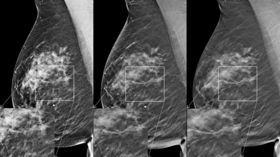
AI competition furthers research on computer-aided detection in breast imaging
A recent AI challenge in the advancement of automated digital breast tomosynthesis (DBT) yielded impressive results that promise to benefit the future of breast cancer detection.
Eight teams were tasked with developing algorithms capable of achieving high sensitivity for lesion detection on DBT. Using thousands of exams in numerous datasets, the teams developed algorithms that yielded excellent detection results, with the highest sensitivity recorded as 0.957 [1].
Details of the competition were shared recently in JAMA, where experts involved in the work suggest that such initiatives could be a driver for research in the field.
“An accurate and robust artificial intelligence algorithm for detecting cancer in digital breast tomosynthesis could significantly improve detection accuracy and reduce healthcare costs worldwide,” corresponding author Nicholas Konz, with the Department of Electrical and Computer Engineering at Duke University, and co-authors note. “The variety of approaches that participants used in this study, alongside their released code and our released tumor detection benchmarking platform, present a starting point for future research in this area.”
For the challenge, teams were first given a large dataset of imaging on healthy individuals to develop and train their algorithms, followed by a smaller dataset to refine their methods. Final rankings were based on how the algorithms performed on an additional dataset containing both normal and cancerous tissue.
In the end, a team from NYU Langone Health (NYU B-Team) recorded the highest sensitivity of 0.957, followed by a team from IBM-Haifa (ZeDus) with 0.926. The mean sensitivity of all submitted algorithms was 0.879.
Teams were instructed to share detailed descriptions and analyses of their algorithms, along with their performance predictions and code for their selected method. The authors suggest that in sharing this data, it could help to “lay the groundwork for the development of clinically relevant computer-assisted diagnosis systems.”
Full details of the competition and its participating teams can be found here.

