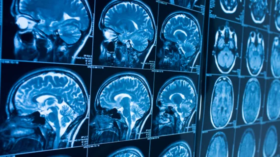
COVID-19 brain MRI findings reveal three distinct neurological patterns
Data establishing a connection between COVID-19 and adverse neurological symptoms continues to grow, with new research released Tuesday showing three main brain MRI patterns in patients with the disease.
The French Society of Neuroradiology examined 37 consecutive patients’ neurological imaging findings from 16 different centers for their study, published June 16 in Radiology. It is the first to characterize a large group of confirmed brain MRI abnormalities, excluding stroke, that are related to the novel virus.
Most patients—who averaged 61 years old—presented with altered consciousness (73%), pathological wakefulness after sedation was ceased (41%), confusion (32%) and agitation (19%).
MRI scans most often revealed signal abnormalities in the medial temporal lobe (43%), an area of the brain related to critical cognitive and emotional functions.
Fewer patients (30%) showed “non-confluent multifocal white matter hyperintense lesions on FLAIR, and diffusion sequences with variable enhancement, with associated hemorrhagic lesions.” This has “rarely” been found in COVID-19 patients, but closely resembles chronic disorders that destroy fatty protective nerve coverings, Stéphane Kremer, with Hospital de Hautepierre, and colleagues wrote.
The third brain MRI pattern was found in 24% of individuals, described as “extensive and isolated white matter microhemorrhaging.” This condition was reported in seven other critically ill patients involved in a separate study, but the authors could not determine the exact physiological process associated with their own findings.
A majority of patients in this cohort had intracerebral hemorrhagic lesions (54%) and a more severe case of COVID-19, the authors noted.
COVID-19’s impact on the central nervous system remains a mystery. One study published last week in the Journal of Alzheimer’s Disease suggested performing baseline MRI prior to discharging patients in order to establish a starting point for evaluating these individuals. That same study proposed a three-stage “NeuroCovid” framework to classify virus-related brain damage.
“Three main neuroradiological patterns could be distinguished, and the presence of hemorrhage was associated with worse clinical status,” the authors wrote. “SARS-CoV-2 RNA was detected in the cerebrospinal fluid in only one patient, and the underlying mechanisms of brain involvement remain unclear,” they noted, adding that imaging and neurological follow-up will allow providers to learn more about these patients.

