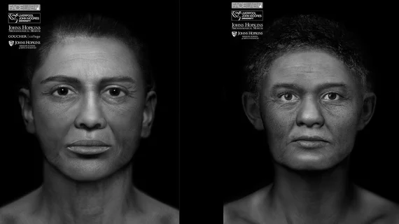
CT imaging of ancient remains creates 3D pictures of Egyptian mummy faces
Two Egyptian mummies pinned unidentifiable by archaeologists have recently been given three dimensional (3D) faces with the help of CT imaging and computer animation thanks to researchers at Johns Hopkins University in Baltimore, according to a report published Oct. 23 by The Baltimore Sun.
The detailed portraits were created in coordination with Face Lab, a research group at Liverpool John Moores University in England that conducts forensic and archaeological research to perform 3D forensic facial reconstruction.
Elliot K. Fishman, PhD, a professor of radiology and the director of diagnostic imaging at Johns Hopkins Hospital, performed the initial CT scan on the bodies of the two ancient figures. The images were given to Face Lab with a blueprint for which to create a 3D representation of each figure. The facial illustrations have been in the works for the past two years, as researchers were “allowing them to take shape at their own pace,” according to the article.
The public is invited to come face-to-face with the 2,300-year-old deceased figures in the exhibit, “Who Am I? Remembering the Dead Through Facial Reconstruction,” currently on display at the Johns Hopkins University Museum of Archaeology in Baltimore.
“That’s one of the opportunities we have with this exhibit — to be able to say, ‘You know what? These people have been with us since the 1880s, and we’re only now able to see them as real people,’” Sanchita Balachandran, the museum’s associate director and the driving force behind the project, told The Baltimore Sun.
See The Baltimore Sun’s entire article below.

