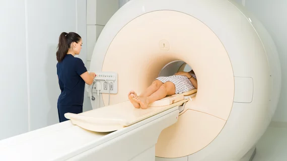
Deep learning-based reconstruction nearly halves spinal MRI acquisition times
Deep learning-based image reconstruction techniques can shave a substantial amount of time from spinal MRI exams, improving both patient comfort and workflow efficiency.
What’s more, the accelerated exam does not come at the expense of image quality, according to new work published in Academic Radiology.
Compared to the standard of care, researchers determined that spinal MRI exam times can be nearly halved when deep learning image reconstruction (DLIR) techniques are deployed. Considering the growing demand for spinal MRIs and the discomfort of the exam for patients struggling with back pain, any time saved on the exam could significantly improve patient care and the workflows of busy imaging departments, authors of the new study suggest.
“Conventional MRI has several drawbacks, including long scanning times which may degrade image quality due to motion artifacts, patient discomfort with increased anxiety,” senior author Shiyuan Liu, MD, with the Department of Radiology at the Second Affiliated Hospital of Naval Medical University in China, and colleagues explain. “Therefore, reducing MRI acquisition time while maintaining high-quality diagnostic images becomes crucial given increasing clinical demand of spinal MRI nowadays.”
For the study, the team compared the imaging of a group of 200 participants who underwent both conventional and accelerated MRI scans for back pain, the latter of which utilized DLIR post-scan. Radiologists evaluated the resultant images’ signal-to-noise ratios (SNR) and contrast-to-noise ratios (CNR), in addition to providing subjective quality and lesion characteristic assessments for the exams.
Using DLIR, scan times were successfully reduced by around 40%. On the accelerated exams, SNR and CNR were significantly higher, and both subjective quality and the frequency of lesion detection were comparable between the two scans. The DLIR exams were found to be interchangeable with the standard of care in detecting different spinal abnormalities, and in some cases, the image quality was better using deep learning recons.
“DL-based models in image reconstruction can overcome the SNR/scan time relationship by applying detail-preserving denoising to accelerate sequences and restore quality to [standard-of-care] levels. Our results revealed that the DL image, compared with SOC, enabled an approximately 40% reduction in overall examination time,” the authors note. “Besides the reduction in scanning time, the image quality of spinal MR images was also improved.”
The prospective, real-world nature of the study is a big strength and highlights the clinical utility of DLIR, the group suggests. However, future studies that focus on similar tools’ utility when exams are acquired on different equipment and magnetic fields are warranted to determine generalizability.
The study abstract is available here.

