
Easy-to-use AI model effectively segments 80 structures on MRI
Researchers have developed a new artificial intelligence model capable of outperforming publicly available segmentation algorithms.
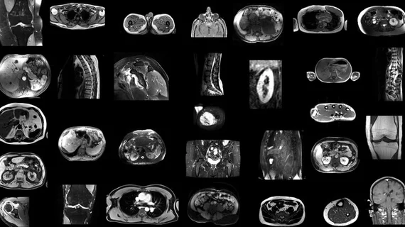
The tool—an nnU-Net model known as TotalSegmentator MRI—provides rapid segmentation of all major organs across all MRI sequences. The open-source model could drastically improve the time providers spend completing manual segmentations, experts involved in its development suggest.
“MRI images have traditionally been manually segmented, which is a time-consuming process that requires intensive effort by radiologists and is subject to inter-reader variability,” Jakob Wasserthal, PhD, a radiology research scientist at University Hospital Basel in Switzerland, said in a news release. “Automated systems can potentially reduce radiologists' workload, minimize human errors and provide more consistent and reproducible results.”
The tool can segment 80 anatomical structures visualized on MRI. It was trained using thousands of MRI and CT scans sourced from routine clinical studies. Its ability to adapt to different datasets and automatically adjust its architecture, preprocessing and training strategies mimics that of a similar model—TotalSegmentator CT, which has more than 300,000 users globally.
Researchers evaluated the model’s performance using Dice scores, which enabled the team to compare its accuracy with that of the radiologists who originally carried out the manual segmentations. The model performed well across all 80 anatomical structures, achieving a mean Dice score of 0.84 on an internal MRI test set. What’s more, this score was significantly higher than those achieved by two publicly available segmentation models currently in use, and is also matched the performance of TotalSegmentator CT.
Experts believe the model could provide significant benefits in treatment planning, disease monitoring and opportunistic screening.
“To our knowledge, our model is the only one that can automatically segment the highest number of structures on MRIs of any sequence,” Wasserthal said. “It’s a tool that helps improve radiologists’ work, makes measurements more precise and enables other measurements to be done that would have taken too much time to do manually.”
Learn more about the team’s work here.

In addition to her background in journalism, Hannah also has patient-facing experience in clinical settings, having spent more than 12 years working as a registered rad tech. She began covering the medical imaging industry for Innovate Healthcare in 2021.