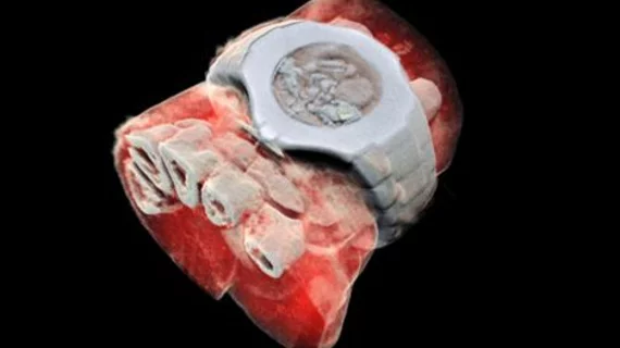
First human imaged with novel 3D color x-ray scanner in New Zealand
A 3D color medical scanner invented by father and son scientists in New Zealand recently imaged its first human subject, according to a news release from the University of Canterbury in New Zealand.
The novel imaging device, called the MARS spectral x-ray scanner, can capture enhanced detail of the body's chemical components—such as fat, water, and disease markers—by measuring the x-ray spectrum to produce images in color. Producing more detailed images than MRI or CT may allow physicians to identify and diagnosis diseases earlier.
Phil Butler, PhD, a physicist at the University of Canterbury, and his son, Anthony Butler, PhD, a radiology professor at the University of Canterbury and the University of Otago in New Zealand, invented the scanner—and Phil Butler happened to be the first person to be scanned by the MARS spectral x-ray scanner, according to the release.
“So far researchers have been using a small version of the MARS scanner to study cancer, bone and joint health and vascular diseases that cause heart attacks and strokes," Anthony Butler said in a prepared statement. "In all of these studies, promising early results suggest that when spectral imaging is routinely used in clinics it will enable more accurate diagnosis and personalization of treatment.”
The duo will soon conduct a clinical trial with orthopedic and rheumatology patients to compare the images produced by the MARS spectral x-ray scanner with current imaging technology.

