New ‘groundbreaking’ imaging device spots early cancers, beats MRI and CT resolution
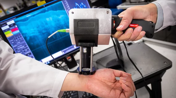
Scientists at Nanyang Technological University in Singapore have created a new handheld medical imaging device that produces higher-resolution scans compared to many modalities and may go on to alter the oncology landscape.
The prototype relies on micro optical-coherence tomography, a new technology that penetrates human tissue and organs, and measures the delay time between the "echo" of light waves as they touch tissue structures. It’s been tested in a few clinical trials thus far and proved to yield images nearly 100-times higher resolution than x-ray, CT and MRI machines.
“This is a groundbreaking technology that could have widespread clinical applications,” said Eng Soo Yap, a consultant hematologist at the National University Hospital in Singapore, who wasn’t involved in the study. “These range from real-time imaging of tissues at a microscopic level to even detecting circulating cancer cells in the blood.”
“All this could lead to early and more accurate detection of cancer,” he added in a statement.
How can micro-OCT accomplish this? The novel device can produce images down to about 1 to 2 micrometers, identifying the very first signs of tumor development within individual cells.
And its proven effective at determining whether colon polyps are malignant or benign. During a preliminary trial at Renmin Hospital of Wuhan University, clinicians used the new imaging device to assess 58 tissue samples taken from growths in patients’ colon or rectum. Performed in real-time, the micro-OCT approach was 95% accurate compared to senior pathologists.
What’s more, the technology is handheld and designed to be used by healthcare workers who aren’t seasoned in imaging or pathology, the authors noted.
“Our device is a fraction of the size of existing machines and produces clear, high resolution images in real-time,” said Liu Linbo, lead researchers and associate professor at NTU.
The entire project is the product of six years of research collaboration between NTU, Harvard Medical School and the University of Alabama. With further improvements currently underway, the team expects the device to eventually move to patients’ bedsides, into homes and more.
"It is our hope that in [the] future doctors might be able to use a device like ours to precisely identify diseases as they develop at the cellular level, in real-time, and in high resolution," Liu said. "Through earlier detection, we believe that patients will receive an earlier diagnosis and if necessary, get treatment faster."

