Inexpensive eye imaging method may help monitor Alzheimer’s progression
Queen’s University Belfast researchers found the eye may be a critical indicator for Alzheimer’s disease (AD) along with a host of other neurodegenerative diseases.
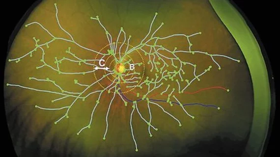
Published in the Journal of Ophthalmic Research, the study showed peripheral retinal imaging, using ultra-wide field (UWF) technology, can be used to visualize changes in the peripheral retina that are associated with these debilitating diseases.
“These exciting research results suggests that our original hypothesis was right and wide field eye imaging could indeed help monitoring disease progression in patients with AD,” said corresponding author, Imre Lengyel, PhD, with Queens’ University’s School of Medicine, Dentistry and Biomedical Sciences in the U.K. in a release.
Lengyel and colleagues took UWF images of 59 patients with AD and 48 healthy participants to determine a baseline. The entire cohort was invited for a follow-up imaging after two years.
One significant finding from the study was that those with Alzheimer’s had a higher than normal appearance of drusen—yellow spots that show up on retinal images in people with AD. Drusen are little deposits of fat, proteins and minerals that, in small numbers, are harmless, but as their numbers increase lead to retinal degeneration, according to the release.
Additionally, the team found those with AD have wider blood vessels near their optic nerve, but these vessels thin faster than seen in the control group near the retinal periphery. This thinning is likely to slow blood flow and hinder nutrient and oxygen flow to the peripheral retina, according to the authors.
Although retinal imaging cannot diagnose AD and other neurodegenerative diseases, co-author Craig Ritchie, a professor of psychiatry of Aging at the University of Edinburgh said the imaging technique could be a simple, quick and cheap way to monitor eye changes and act as a tool to easily measure disease progression in the brain.
“Our research team … was able to identify early markers in people many years before dementia develops,” Ritchie said in the statement. “We have opened a window to identify high risk groups who may benefit from specific prevention advice.”

