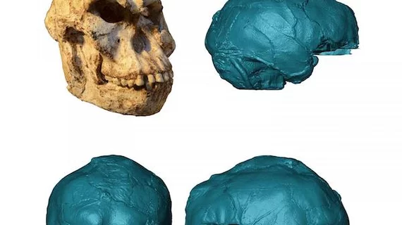
Micro-CT used to reconstruct 3.6M year old brain
What does the brain of 3.67 million year old hominin known as Little Foot look like? Thanks to Micro-CT scans of the ancient fossil, researchers reconstructed its brain and are learning more about the organ’s early evolution.
Amélie Beaudet, PhD, of Wits University in Johannesburg, South Africa and colleagues “virtually extracted” brain scans of the ancient human relative and published their findings in the Journal of Human Evolution. The images unveiled impressions from when the skull housed Little Foot’s brain, its shape and vessels.
It also revealed an asymmetrical brain and expanded visual cortex, suggesting these features were shared between the last common ancestor of hominins and other great apes, according to the authors.
Examining the scans, Beaudet et al. also found a more complex vascular system than once thought. This finding, they noted, supports the theory that the endocranial vascular system in Australopithecus was more similar to modern humans than its younger relatives.
"This would mean that even if Little Foot's brain was different from us, the vascular system that allows for blood flow (which brings oxygen) and may control temperature in the brain—both essential aspects for evolving a large and complex brain—were possibly already present at that time," Beaudet said in a Wits press release.
Overall, the findings point to a more complex evolution in the brain structures of younger hominins experienced more than three million years ago. Perhaps, the authors wrote, this was a response to increasing environmental pressures.
"Such environmental changes could also potentially have encouraged more complex social interaction, which is driven by structures in the brain," Beaudet said in the statement.

