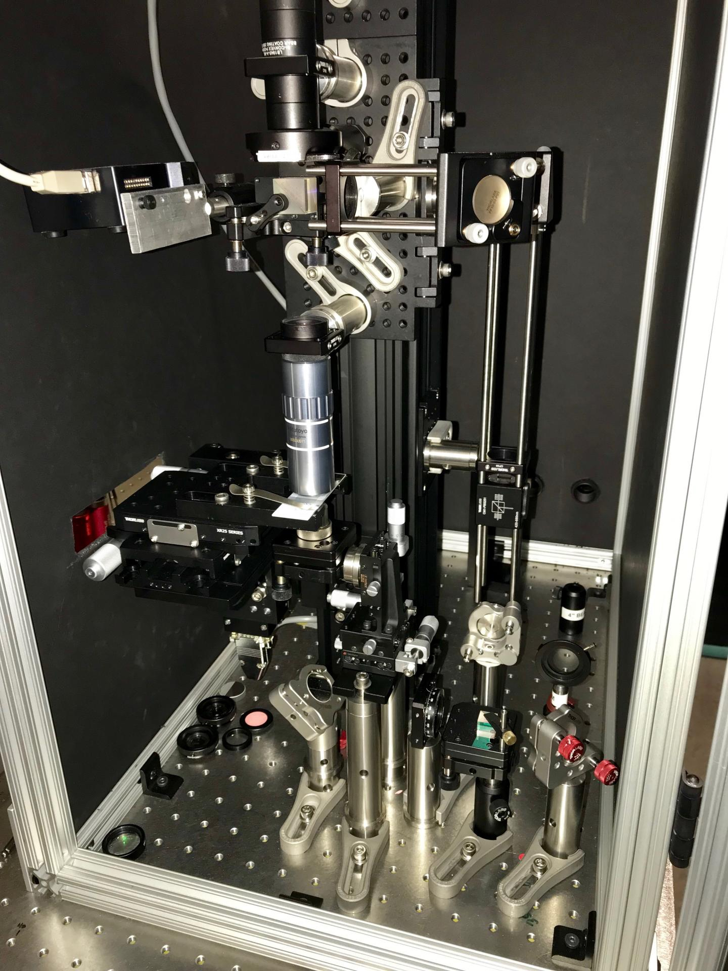
New optical tomography microscope bolsters the power of 3D imaging
A novel three-dimensional imaging method created through a multi-institutional effort has great potential for diagnosing cancer and other diseases.
Individuals from Colorado State University and the University of Illinois at Urbana-Champaign teamed up to develop the 3D visualization technique referred to as harmonic optical tomography (HOT). The process harnesses holographic information, such as the intensity and phase delay of light, to create 3D images.
"This new type of tomographic imaging could prove to be very valuable for a wide range of studies that currently rely on two-dimensional images to understand collagen fiber orientation, which has been shown to be prognostic for a number of types of cancer,” Jeff Field, director of the Microscopy Core Facility at CSU, remarked in a statement.

Where this HOT imaging development truly differentiates itself, is in the custom, high-powered laser designed by graduate students at the Colorado institution.
The researchers tested their model on two samples, a manufactured crystal and a biological sample of muscle tissue. HOT imaging was “useful” for looking at both objects, which are typically difficult to study using conventional imaging, the team explained June 1 in Nature Photonics.
"Using this technique, we can use the orientation of the collagen fibers as a reporter of how aggressive the cancer is,” said Gabriel Popescu, professor of electrical and computer engineering at the University of Illinois. He added that collagen is the most prevalent protein in humans and is useful in altering the frequency of light required during HOT imaging.
The National Science Foundation and National Institutes of Health each contributed funding for this research.

