
MRI results affected by movement? MIT researchers have an AI-powered solution
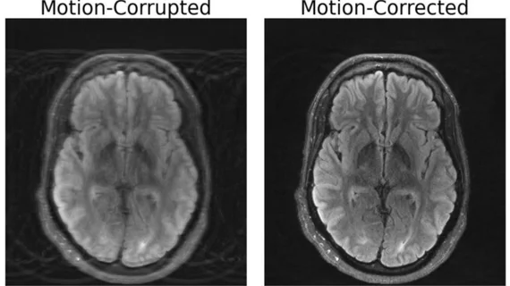
If a patient can’t stay still, technology may now be able to compensate. Researchers at the Massachusetts Institute of Technology (MIT) have unveiled a deep learning model capable of addressing the persistent challenge of motion artifacts in magnetic resonance imaging (MRI) scans. The innovation holds the potential to greatly improve diagnostic accuracy, particularly for patients with neurological disorders.
The paper that initially detailed the discovery, “Data Consistent Deep Rigid MRI Motion Correction,” was recently awarded best oral presentation at the Medical Imaging with Deep Learning conference in Nashville back in July.[1]
During the presentation, lead author Nalini Singh, an Abdul Latif Jameel Clinic for Machine Learning in Health (Jameel Clinic)-affiliated PhD student in the Harvard-MIT Program in Health Sciences and Technology detailed how researchers computationally constructed a motion-free image from data with artifacts using existing MRI technology.
The significance of this integrated method lies in its ability to guarantee coherence between the generated image and the real measurements, as any divergence can lead the model to generate "hallucinations” – fabricated images that can seem authentic, but lack precision in their physical and spatial representation. Such inaccuracies could potentially exacerbate the impact on diagnostic outcomes.
“Our aim was to combine physics-based modeling and deep learning to get the best of both worlds,” Singh explained in a statement.
MRI scans are known for their superior soft tissue contrast compared to other imaging modalities like X-rays or CT scans. However, their sensitivity to even the slightest patient movement during scans often leads to image artifacts, risking misdiagnoses and redundant scans. Patients, including those with neurological disorders, are sometimes asked to limit their breathing or are even anesthetized to minimize movement – but such measures are not always feasible.
This breakthrough could have far-reaching implications for medical diagnostics, particularly for patients with conditions like Alzheimer's or Parkinson's disease, which involve involuntary movements. Singh anticipates that her research can be extended to address more complex types of motion, including fetal MRI, which is challenged by unpredictable and rapid movements.
Fixing motion artifacts may also bring significant cost savings. According to a study from the University of Washington Department of Radiology, motion affects around 15% of brain MRIs. The cost of artifacts is substantial, leading to approximately $115,000 in annual hospital expenditures per scanner due to repeated scans required to obtain usable images.
"Not only is it excellent research work, but I believe these methods will be used in all kinds of clinical cases: children and older folks who can't sit still in the scanner, pathologies which induce motion, studies of moving tissue, even healthy patients will move in the magnet,” Daniel Moyer, an assistant professor of computer science at Vanderbilt University, said in the same MIT statement. “In the future, I think that it likely will be standard practice to process images with something directly descended from this research.”

Chad is an award-winning writer and editor with over 15 years of experience working in media. He has a decade-long professional background in healthcare, working as a writer and in public relations.