MRI scan data predicts preterm infants at risk of developmental brain issues
Magnetic resonance imaging scan data can help predict which premature infants are most likely to face developmental issues, new research out of Cincinnati Children’s Hospital Medical Center suggests.
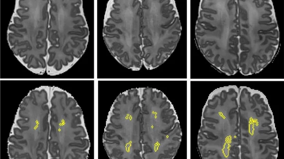
The team detailed its novel software tool that quantifies diffuse white matter abnormality on MRI scans Sept. 28 in Scientific Reports. It revealed DWMA volume to be an independent predictor of motor development deficits, such as cerebral palsy.
Lead researcher Nehal Parikh, DO, MS, a neonatologist at Cincinnati Children’s, said the findings challenge conventional wisdom on the topic.
"While most researchers and doctors have concluded that DWMA is not pathologic, our novel studies are concluding otherwise," Parikh added in a statement on Wednesday. "Most studies have diagnosed DWMA qualitatively based on visual readings from radiologists. These subjective diagnoses have been unreliable and therefore have not been significantly associated with neurodevelopmental disorders."
For their investigation, the researchers prospectively enrolled 110 preterm infants (born at 31 weeks) from four neonatal intensive care units. These NICUs care for nearly 80% of preterm babies in the Columbus, Ohio, area, Parikh et al. noted.
After the automated software tool analyzed structural brain MRIs taken from all infants, DWMA volume was a strong predictor of motor development issues at three years old. Currently, many of these children aren’t diagnosed with these disorders until they’re five.
In the near future, Parikh’s team will complete its large study called the Cincinnati Infant Neurodevelopment Early Prediction Study (CINEPS) to further validate their findings.
"We plan to work with MRI manufacturers to incorporate our software onto their systems in order to provide objective diagnoses of DWMA at the point of care," Parikh concluded. "This advance will permit accurate parental counseling and early risk stratification to enable targeted early intervention therapies."

