New AI technology developments consistently recognize medical image anomalies
A study published in IEEE Access cites the advancements involving the use of AI in medical imaging. Scientists from Skoltech, Philips Research and Goethe University Frankfurt have taken an innovative approach in the pursuit AI implementation by deploying new methods in the setup of AI models.
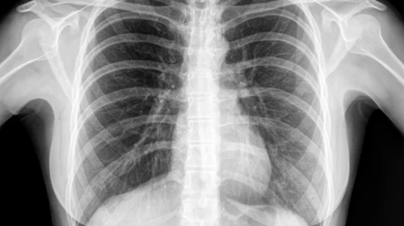
For this study, researchers used a database of chest x-rays and breast cancer histology microscopy images. Researchers used an autoencoder approach and enacted what the study refers to as “progressive growing training,” which would allow for a small amount of image anomalies to be included in the model setup, with more added on a regular basis. In previous models, no anomalies were present. “We showed some abnormal images to the network to unleash the arsenal of weakly supervised methods, and it helped a lot. Even just one anomalous scan for every 200 normal ones goes a long way.” said Skoltech Professor Dmitry Dylov, senior author of the study, and head of the Institute’s Computational Imaging Group, in a press release sent by Skoltech.
According to the study, the new method outperformed previous methods in every case considered. The goal of this new method is for the model to be able to “perceive” the data just like the radiologist would, looking for any anomalies beyond what a “normal” baseline is typically. By simplifying the approach, this new method also presents a potential building block for other researchers to reference and build upon while keeping the foundation of the research consistent.
Realistically, the “normal” scans a radiologist views far outnumber the “abnormal” ones. The hope of this study is that by training AI to recognize small scale anomalies, radiologists and specialists can eliminate the need to review unproblematic imaging, thus eliminating some of the burdensome work required to keep up with patient demand.
You can read the full study for free here.

