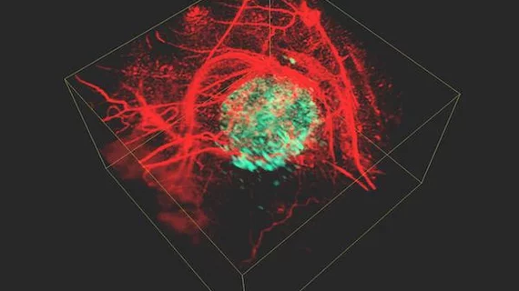
New photoacoustic imaging method offers 3D look at cancer cells
Researchers have developed a new method for creating three-dimensional (3D) images of cells that utilizes photoacoustic imaging and a phytochrome protein to provide in vivo looks at tumor growth.
The findings, published in Communications Physics, may allow for a more thorough understanding of cancer cells in the body, according to the researchers with Martin-Luther Universität (MLU) Halle-Wittenberg in Germany.
"Our aim is to visualize cancer cells inside the living body to find out how they function, how they spread and how they react to new therapies," said Jan Laufer, from MLU, in a statement.
Photoacoustic imaging can produce high-resolution images of cells at depths optical microscopy cannot. Researchers used a method based on reversibly photoswitchable phytochrome-based reporter protein (AGP1) along with dual-wavelength interleaved image acquisition to produce the 3D images.
Laufer explained the method more plainly in the statement. Scientists introduce an individual gene into the genome of the cancer cells. That gene produces a phytochrome protein which acts as a light sensor. Short pulses of light at two different wavelengths illuminate the tissue, which pulses are absorbed inside the body and released back out as ultrasonic waves.
Once outside the organism, these waves can be measured, with the data used to generate two images of the body’s interior. The high-resolution 3D image is calculated by measuring the difference between the two.
“This method represents a powerful new approach to studying cellular and genetic processes which, due to its experimental simplicity, can be implemented in a wide range of existing photoacoustic imaging platforms,” wrote Laufer and colleagues.

