Harvard researchers’ novel molecular imaging technique creates brain 'cellular atlas'
Harvard University researchers used novel molecular imaging technology to make a first-of-its-kind “cellular atlas,” covering a key area in the brain to help study the genetic makeup and function of cells, according to research published online Nov. 1 in Science.
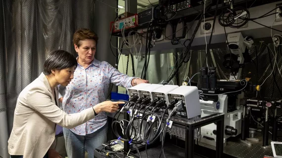
A team led by Catherine Dulac, PhD, and Xiaowei Zhuang, PhD, of Harvard used their imaging technology called Multiplexed Error-Robust Fluorescence In-Situ Hybridization (MERFISH) to examine more than one million cells in the brain’s hypothalamus—the region of the brain responsible for thirst, feeding, sleep and social behaviors like parenting and reproduction.
“This [study] gives us a granular view of the cellular, molecular and functional organization of the brain—nobody had combined those three views before,” Dulac said in a prepared statement. “This work in itself is a breakthrough because we now understand several behaviors in ways that we never did before, but it’s also a breakthrough because this technology can be used anywhere in the brain for any type of function.”
The study was primarily designed to address the problem researchers have when studying components of cells. The process usually involves disassociating cells from the tissue, which, in turn, can lose information about how the cells were organized in the tissue and what other cells surrounded them.
“If you really want to understand the brain, you need the spatial context, because the brain is not like the liver or other organs, where the cells are organized in a symmetrical way,” Dulac said. “The brain is unusual in that it has this topological arrangement of neurons…so we want to be able to look at a section of the brain and see what cells are there, but also where they are and what types of cells are surrounding them.”
Using MERFISH imaging in mice, the researchers imaged all molecules functioning in cells within the brain’s hypothalamus. MERISH works by assigning “genetic barcodes” to the cell’s RNAs. Through multiple rounds of imaging, the barcodes are read out to determine the cells identity and source of origin, according to the researchers.
The method allowed the researchers to image and distinguish thousands of different RNAs in just 10 rounds of imaging. The team also selected a subset of barcodes that could only be misread if multiple errors occurred at once, which ultimately helped reduce the risk of misidentifying a gene.
“One of the main applications that we invented MERFISH for is to identify cell types in situ because different cell types have different gene-expression profiles. Hence, these gene-expression profiles provide a quantitative and systematic way for cell-type identification,” Zhuang said. “And because we can do this in intact tissues by MERFISH imaging, we can provide the spatial organization of these cell types, too.”
Additionally, Dulac, Zhuang and colleagues combined MERFISH imaging with single-cell sequencing to gather unbiased quantification of gene expression profiles for the cells. MERFISH was then used to simultaneously image more than 150 genes in the hypothalamus to create a spatial map of the cell's location. The imaging technique also let the researchers identify 70 different types of neurons.
“You can see which neurons are neighboring each other…and not only that, but because our images are molecular, you can identify how these cells are communicating with each other,” Zhuang said. “Moreover, because MERFISH imaging has a very high sensitivity, we were able to identify lowly expressed genes that are critical to cell function.”
With these findings, the researchers linked together specific cells with specific behaviors and ended up with a solution in the form of a gene called c-Fos, also known as an “immediate early” gene, according to the researchers. The gene is increased during neural activity, so researchers can track which cells show increases in the gene and which cells are activated during certain behaviors.
“The identification of marker gene combinations and spatial locations defining the neuronal populations in the preoptic region provides necessary tools for the precise targeting and perturbation of these neurons, thus enabling future functional studies,” the researchers wrote. “As an imaging-based approach, we envision that MERFISH can be combined with diverse imaging methods for anatomical tracing and functional interrogation to provide insights into how distinct cell types communicate to form functional circuits in the healthy and diseased brain, as well as in other tissues.”

