Novel quantum dots may allow for 3D cell imaging
Researchers from the University of Illinois at Urbana-Champaign and the Mayo Clinic in Rochester, Minnesota teamed up to create a new molecular probe which can measure and count RNA in cells and tissue as well as traditional dyes, according to a new study in Nature Communications.
The probe is taken from a conventional fluorescence in situ hybridization (FISH) technique, but uses compact quantum dots to illuminate molecules instead of fluorescent dyes. Made of zinc, selenium, cadmium and mercury alloy, the dots can emit color regardless of the size of the particle—unlike most quantum dots.
Additionally, their new discovery can overcome the limitations of dyes used during the FISH process which can quickly deteriorate and aren’t suitable for three-dimensional (3D) imaging, according to University of Illinois news story.
"By replacing dyes with quantum dots, there are no stability issues whatsoever and we can count numerous RNAs with higher fidelity than before," said Andrew Smith, an associate professor of Bioengineering and member of the research team, in the story. "Moreover, we uncovered a fundamental limit to the size of a molecular label in cells, revealing new design rules for analysis in cells."
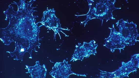
In testing HeLa cells and prostate cancer cells, the team found dye-based FISH cell counts declined in minutes, but their new quantum dot method allowed RNA to be counted for more than 10 minutes and made it possible to perform 3D cell imaging.
"This is important because images of cells and tissues are acquired slice-by-slice in sequence, so later slices that are labeled with dyes are depleted before they can be imaged," Smith added.

