Junior and senior radiologists benefit from AI assistance when identifying pulmonary nodules
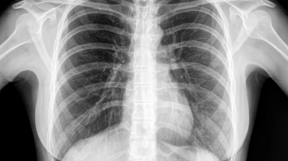
Radiologists benefit from AI-assistance when identifying pulmonary nodules on chest radiographs regardless of their experience level, according to recent research. Since most early lung cancers initially present as pulmonary nodules, these new findings could benefit patients.
The new study published in JAMA Network Open compared radiologists' interpretations of chest radiographs with AI and without assistance. It also went one step further by presenting data on varying degrees of detection difficulty and the experience levels of the interpreting radiologists.
“Although several recent studies have examined how AI may improve the performance of radiologists in detection of pulmonary nodules on CXRs, they are limited in their scope owing to the lack of multisite readers with different levels of clinical experience, detection difficulties, and settings,” corresponding author, Fatemeh Homayounieh, MD, with the Department of Radiology at Massachusetts General Hospital and Harvard Medical School, and co-authors explained.
The study included 100 chest radiographs obtained between 2000-2010. The novel AI algorithm, AI Rad Companion Chest X-ray, was implemented alongside two thoracic radiologists (14 and 16 years of experience) who established ground truth. Nine test radiologists independently reviewed the images using AI and then without its help.
Though both groups of readers benefited from AI assistance, the junior radiologists saw more marked improvement in their interpretations. On average, the detection accuracy of the junior radiologists increased by 6.4%, and their false positive rate also improved by 5.6%. Both groups of readers experienced enhanced sensitivity for pulmonary nodule detection. But for the junior radiologists, the AI-assisted reads improved their sensitivity and specificity by an average of 10.4%.
“Given the high miss rate and importance of identifying pulmonary nodules on chest radiographs, AI algorithms such as the one assessed in our study and those tested in prior studies can play an important role in improving quality and diagnostic value of radiographs in patients with pulmonary nodules, not least in helping to reconcile differences in radiographic equipment from different vendors,” the doctors wrote.
You can view the detailed research in JAMA Network Open.

