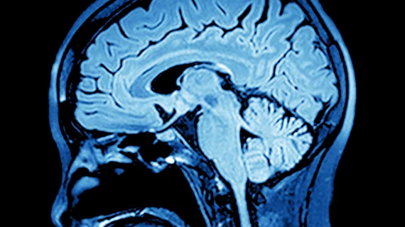
Researcher awarded $600K grant to study MRI-based ‘GPS maps’ for brain tumors
A researcher from Case Western Reserve University has been awarded a grant to test if MRI-based biomarkers can improve how the field manages brain tumors.
The V Foundation—named after former basketball coach Jim Valvano, who died from cancer— awarded Pallavi Tiwari, assistant professor of biomedical engineering at the Cleveland, Ohio-based institution, a three-year, $600,000 grant to bring her “GPS maps” to clinical testing. So far, the approach has done well in retrospective cases, and Tiwari is hopeful those results with translate to the clinic.
“We develop these cool technologies and we get excited about 90% accuracy, but it doesn’t mean anything until you can prove it in clinical trials—because that’s when patients can begin to get the benefits,” she said in a news release. “That’s why this is so exciting for me.”
Traditionally, post-operative MRI is used to evaluate cancer recurrence in patients with brain tumors, however, both the effects of radiation and return of cancer look the same on MRI scans, Tiwari noted. This, coupled with the high cost of invasive brain biopsy—another method to confirm cancer—prompted her to develop a new approach.
That concept derives image-based biomarkers from MRI scans. And it has been about 92% accurate in differentiating cancer recurrence from radiation effects in about 200 cases
“We’ll use the routinely acquired MRI scans, feed them into our computational algorithm and create a GPS map that the surgeon can use—one that will have a ‘heat map’ of hot-spots for cancer to guide him or her in finding the correct biopsy site within the lesion,” Tiwari said.
Clinical trials are expected to kick off in the last year of the grant in 2021 or early 2022, according to Tiwari. Success in these trials may be key to saving patients and money, she added.

