
Are synthetic contrast-enhanced breast MRI images as good as the real thing?
Generating simulated contrast-enhanced images from a non-contrast breast MRI could increase accessibility of the advanced imaging with comparable diagnostic results to real contrast-enhanced MRI scans.
The simulated images offer clinicians additional tumor visualization without patients having to undergo additional imaging that requires the administration of gadolinium-based contrast agents (GBCA). Although GBCAs are considered safe, their use can still be inhibited by a number of factors, which can, in turn, limit the accessibility of contrast-enhanced breast MRI—the most sensitive test for breast cancer detection—for some women, according to the authors of a new paper published in Radiology.
“The administration of gadolinium-based contrast agents requires intravenous access and physician monitoring, which increases the cost and length of breast MRI examinations,” corresponding author Maggie Chung of the University of California San Francisco and co-authors explained. “The need to administer gadolinium-based contrast agents also limits the use of MRI in the small subsets of patients with severely impaired renal function and in pregnant patients.”
The authors explained how synthesizing contrast-enhanced imaging from pre-contrast inputs using deep learning has been shown to be beneficial in overcoming obstacles involved in the use of GBCAs. Prior research focused on imaging of the brain, but Chung and colleagues sought to understand whether the positive results from those studies could translate into clinical utility for breast MRI exams.
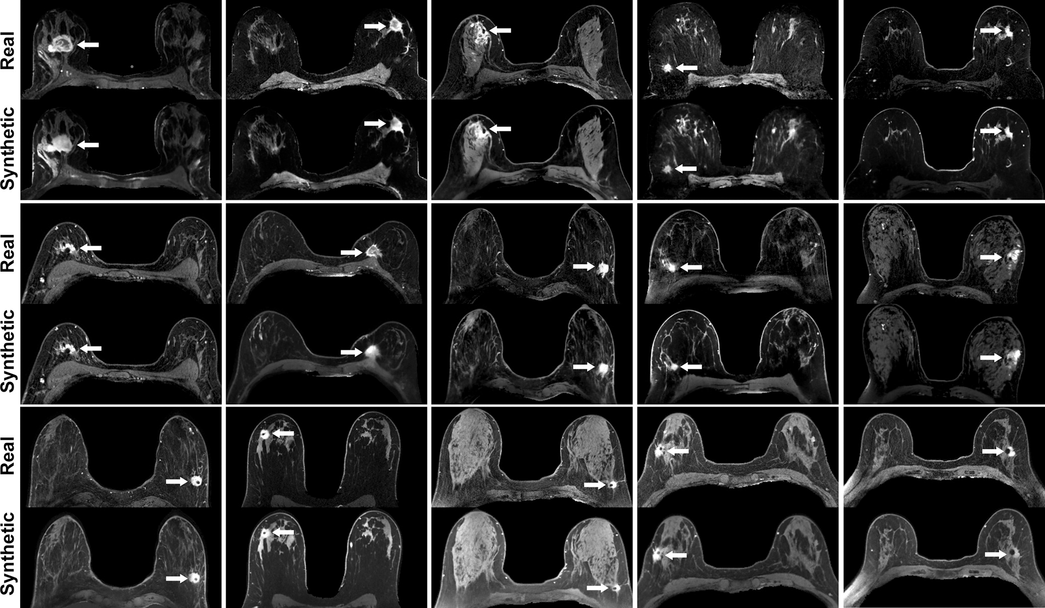
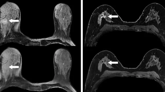
Real versus simulated (ie, synthetic) contrast-enhanced T1-weighted axial breast MRI scans of patients with invasive breast cancer. Pairs of real and simulated contrast-enhanced breast MRI scans from 15 patients with invasive breast cancer (arrows). Intrathoracic and extramammary structures were masked in all images. Image courtesy of RSNA.
For their study, the experts used five pre-contrast sequences to develop a three-dimensional, fully convolutional deep neural network to simulate contrast-enhanced T1-weighted breast MRI scans. The resultant simulated images were then compared to the real contrast-enhanced breast MRI exams of 96 women with invasive breast cancer. Four breast radiologists, blinded to which images were authentic and synthetic, graded them for quality, presence of tumor enhancement and mass size.
According to the readers, the synthetic images were assessed as having the appearance of a real MRI with tumor enhancement. The majority of the synthetic MRI exams (84 out of 88) were deemed to be of diagnostic quality and were similar to the real scans in terms of enhancing tumor overlap.
The authors noted that synthetic contrast-enhanced images are in no way a replacement for real images, but that they could “extend the benefits and accessibility of breast MRI by reducing the necessity of contrast enhancement for tumor visualization.”
The study abstract is available here.

In addition to her background in journalism, Hannah also has patient-facing experience in clinical settings, having spent more than 12 years working as a registered rad tech. She began covering the medical imaging industry for Innovate Healthcare in 2021.